A seed looks like a parcel ready to leave: a small object that can be carried away, buried and eventually opened by a growing plant. But before that departure, reproduction has to happen inside the package. Protection creates a problem of access. How does the male contribution reach something the parent plant has begun to enclose?
Devonian fossils make that question unusually tangible. Their reproductive structures have lobes, projecting tips and chambers whose arrangement can be compared across plants. The origin of the seed becomes a history of these working parts, with some much easier to recognize than others.
Keeping the next generation close
In the seed habit, a large reproductive spore—the megaspore—stays inside the tissue that produced it. One functional megaspore supports the female, egg-producing stage of the life cycle. An outer covering, the integument, surrounds that tissue. Pollen supplies the male contribution; after fertilization, the ovule can become a seed containing an embryo. A seed therefore includes tissues belonging to different generations, assembled before dispersal.[1]
That sequence changes what must travel. The female reproductive stage can develop in place while pollen arrives from elsewhere. Retention and enclosure are consequently only part of the evolutionary story. An enclosing structure also needs a way for reproduction to proceed across its boundary.
The earliest examples need not resemble a smooth, sealed kernel. Late Devonian reproductive organs often had coverings divided into free or partly joined lobes. They lacked the neatly defined opening through the integument, called a micropyle, familiar from later ovules. Instead, specialized tissue at the top of the inner spore-bearing organ handled pollen reception.[4]
A projecting tip in Belgium
In 2004, Philippe Gerrienne and colleagues described Runcaria heinzelinii as a possible window into an earlier stage. From the Givetian of Belgium, around 385 million years ago, it preceded well-established Late Devonian seeds by roughly 20 million years. They interpreted its small reproductive structure as an integument surrounding a megasporangium—the organ that would contain a megaspore—with an additional outer cupule. A long extension rose above the surrounding lobes. The team proposed that its exposed tip intercepted airborne pollen.[3]
The material came from a sandstone slab collected at Ronquières, in the Bois de Bordeaux Formation. A subsequent account described hundreds of these structures, each only several millimetres long. Yet abundance did not resolve the essential interior: neither a megaspore nor the female reproductive tissue could be identified confidently. No opening was observed at the tip, and no convincing pollen chamber was recognized below it.[2]
An exposed projection is suggestive; it is not a preserved pollination event. If the tip caught pollen, another mechanism still had to connect that arrival with fertilization deeper inside. The researchers considered possibilities including local dissolution of cells and a tip that had closed after pollination. These remained hypotheses.[2]
The distinction matters because the fossil's age makes an evolutionary sequence so tempting. Put Runcaria before later seeds on a page and the eye supplies an arrow. The anatomy must earn that arrow independently.
Elkinsia and the receiving chamber
By the Late Devonian, Elkinsia polymorpha supplies a more elaborate comparison. Its reproductive anatomy included an inlet above a pollen chamber, part of the arrangement traditionally called hydrasperman reproduction. A central column was interpreted as helping seal the chamber after pollen reception. The apparatus belonged to the inner organ itself.[2]
Meanwhile, the integument remained partly open. In comparisons of early ovules, Elkinsia falls between forms with entirely separate covering lobes and forms with much more extensive fusion. Pollen reception and the joining of those lobes were distinguishable anatomical features. One should not be used as a shortcut for reconstructing the other.[4]
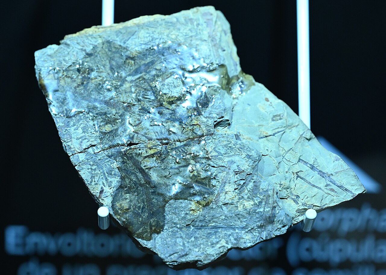
The museum fossil pictured here comes from West Virginia; it is Elkinsia, not the Belgian Runcaria material.[7] The photograph introduces the physical remains, while the anatomical comparison depends on published descriptions of the reproductive organs. A fossil on display and a reconstructed reproductive system offer different scales of access to the same extinct plant.
Nor does “seed fern,” the traditional label applied to Elkinsia and many other extinct plants, identify one tidy lineage of modern ferns acquiring seeds. Paleobotanists have used the term for a diverse assemblage of seed-bearing plants, often with fern-like foliage. Their evolutionary relationships require more than that resemblance.[6]
The sequence remains open
A 2026 reassessment by Richard Bateman, Alan Spencer and Jason Hilton makes the uncertainty around Runcaria sharper. The authors question whether its fossils establish a single retained megaspore, and even whether the source organism was a vascular land plant. They also find the proposed route from the long tip to the presumed female tissue difficult to explain. Its compression preservation kept it outside their analysis of three-dimensionally preserved seeds.[1]
That critique challenges a particular fossil's position. It does not erase the later reproductive organs or turn their anatomical differences into guesswork. We can compare an exposed tip, a receiving chamber and a partly fused covering without claiming to have identified every ancestor connecting them.
During the Carboniferous, ovules diversified further in shape and size, and the integument took on a larger role. Fossils with preserved embryos also enter the record then, adding evidence of the generation inside the enclosure.[5]
The small Devonian structures are compelling because they let us inspect reproduction before the familiar package became familiar. Their open lobes and specialized tips preserve several ways of managing the same difficulty: holding something close while allowing something else to arrive.
Sources
- Richard M. Bateman, Alan R. T. Spencer and Jason Hilton, “Early evolutionary history of the seed,” Biological Reviews, 2026—seed anatomy and the explicit reassessment of Runcaria's affinities and preservation.
- Philippe Gerrienne, Brigitte Meyer-Berthaud and Muriel Fairon-Demaret, “The significance of Runcaria (Middle Devonian, Belgium) in the evolution of seed plants,” Carnets de Géologie, 2005—locality, specimens, missing internal evidence and competing pollination interpretations.
- Philippe Gerrienne et al., “Runcaria, a Middle Devonian Seed Plant Precursor,” Science, 2004—original seed-precursor interpretation and age; author-deposited institutional record and abstract.
- Cyrille Prestianni, “Early diversification of seeds and seed-like structures,” Carnets de Géologie, 2005—pollen-receiving anatomy and the range of integument fusion in Devonian ovules.
- Brigitte Meyer-Berthaud, Philippe Gerrienne and Cyrille Prestianni, “What is a seed?—3. How did we get there? Palaeobotany sheds light on the emergence of seed,” Botany Letters, 2018—institutional record and abstract summarizing Carboniferous diversification and embryo evidence.
- William A. DiMichele, Tom L. Phillips and Hermann W. Pfefferkorn, “Paleoecology of Late Paleozoic pteridosperms from tropical Euramerica,” Journal of the Torrey Botanical Society, 2006—Elkinsia and the diversity encompassed by “seed ferns”; Smithsonian-hosted paper.
- Krzysztof Ziarnek, Kenraiz, “Elkinsia polymorpha kz01.jpg,” photographed 20 March 2024—West Virginia fossil in the Museo de Paleobotánica Roberto Wagner, Córdoba; photograph and provenance, CC BY-SA 4.0.