Inside the mare's sprawling skeleton, a smaller set of bones remains gathered together. This is Eurohippus messelensis, an early horse relative excavated at Messel in Germany in 2000. The cluster belongs to her unborn foal.[1] The photograph invites an immediate recognition of pregnancy. Understanding what surrounds those bones takes a slower kind of looking.
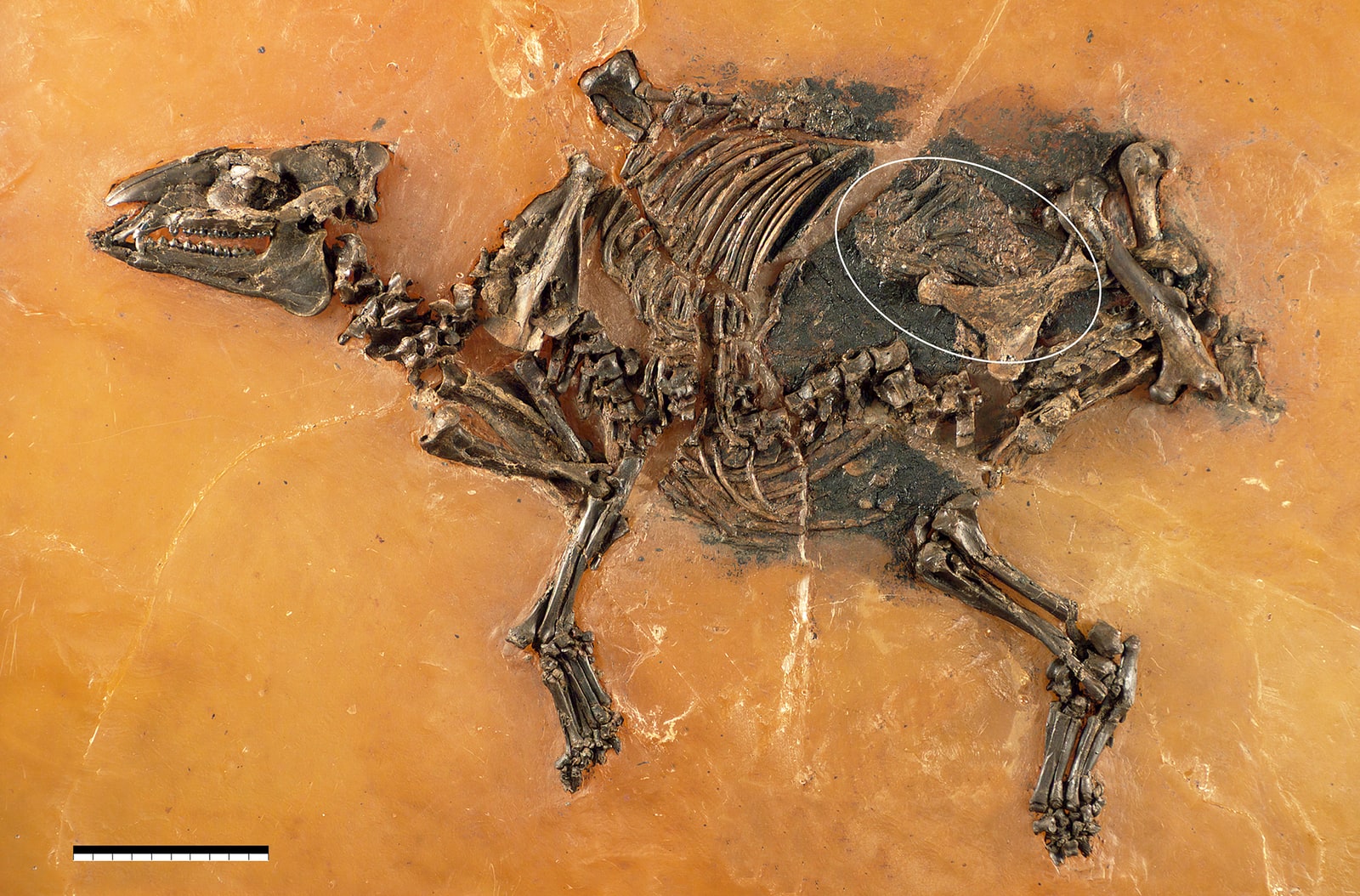
Messel's oil shale records an Eocene environment approximately 47–48 million years old. UNESCO's account emphasizes the unusual range of material preserved there: feathers, hair, skin and stomach contents accompany articulated skeletons.[2] That richness changes the questions a fossil can answer. Teeth and limbs describe an animal's equipment; a pregnancy can reveal a temporary arrangement inside a particular body. Here, the scientific subject is a moment in a life.
The fetal bones are compressed and difficult to identify from the exposed surface. In their 2015 description, Jens Lorenz Franzen, Christine Aurich and Jörg Habersetzer used high-resolution micro-X-rays to distinguish them. Much of the skeleton behind the skull remains connected. Bone development and milk teeth suggested late gestation, while the forelimbs had not yet extended toward the birth canal. The authors interpreted the posture as normal before labour and considered a fatal birth complication unlikely.[1]
The distinction matters emotionally as well as scientifically. A mother and fetus can easily become a story about a disastrous delivery. The interpretation here instead leaves the cause of death unresolved. An intimate association is powerful evidence, but it cannot supply every event that preceded burial.
Around the fetus, the evidence changes character. Franzen's 2017 follow-up described a yellowish mineral crust with fine wrinkles, interpreted as the uteroplacenta: the pregnant uterus together with its associated placental membrane. Microscopy revealed mineralized bacterial forms. The tissue's shape had been recorded through decay and mineralization; the specimen does not contain an intact, preserved womb in the everyday sense.[4]
A further structure runs between the fetal region and the mother's lower backbone. Its position and direction led the researchers to identify a broad uterine ligament, the supporting connection between uterus and body. They compared it with living mares and considered an abdominal-muscle interpretation anatomically inconsistent. Wrinkles alone would be less persuasive than this combination of surface texture, location and attachment.[4]
There is a second history inside that readable surface: the work of preparation. Senckenberg explains that Messel's water-rich shale crumbles when it dries. Preparators expose one side of a vertebrate fossil, support it in artificial resin, and then work from the other side. Fine needles and a stereomicroscope allow delicate structures to be uncovered over days or weeks.[3]
For a reader looking at the photograph, that procedure changes the meaning of a smooth background. The fossil has passed through an intervention designed to keep its parts together. The relationship between neighbouring bones must survive the removal of the material that held them. Preparation therefore belongs in the explanation of how this object can be studied, alongside excavation and imaging.[3]
Franzen's follow-up makes the stakes unusually concrete. In another pregnant Messel mare, a preparator had cut through an unrecognized uteroplacenta and exposed the supporting polyester beneath. Elsewhere on that specimen, a thin layer could represent either fetal skin or an enclosing membrane; its identity remained open.[4] Recognition can arrive after preparation, and exceptional preservation does not make every surviving layer self-explanatory.
This is why the most rewarding way to look at the mare is to keep changing scale. First comes the whole animal, then the small gathering inside its abdomen, then the faint structures around that gathering. Each step asks a different question: what is present, how are the parts related, and what process made them visible? The fossil's intimacy survives that scrutiny. Indeed, it becomes more affecting when the apparent simplicity of the photograph gives way to the patient work required to read it.
Sources
- Jens Lorenz Franzen, Christine Aurich and Jörg Habersetzer, “Description of a Well Preserved Fetus of the European Eocene Equoid Eurohippus messelensis,” PLOS ONE (2015) — specimen, imaging, fetal development and photograph, figure 2.
- UNESCO World Heritage Centre, “Messel Pit Fossil Site” — geological age and exceptional preservation.
- Senckenberg Nature Research, “Location Messel,” Excavation and Preparation — shale instability, resin transfer and preparation practice.
- Jens Lorenz Franzen, “Report on the discovery of fossil mares with preserved uteroplacenta from the Eocene of Germany,” Fossil Imprint 73 (2017), 67–75 — mineralized tissues, anatomical comparisons and preparation history.