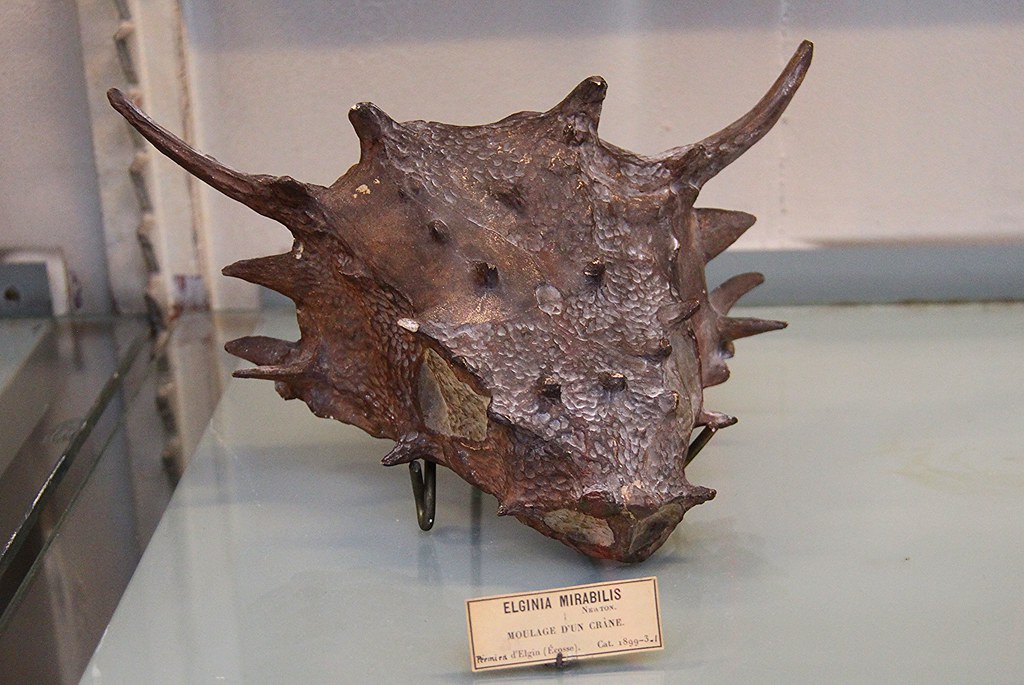
The head spreads outward as though every edge has found a way to become a point. Long horns sweep from the back corners; smaller projections crowd the cheeks and eye sockets. In the Paris museum photograph, the brown skull rests above a label identifying a cast. That small word is particularly appropriate for Elginia: much of what scientists know about this animal began with the shape of bone that had disappeared.[1][2]
Elginia mirabilis was a pareiasaur, a member of an extinct group of reptiles that included substantial plant-eaters. Its Scottish fossils come from the late Permian Cutties Hillock Sandstone near Elgin. The animal belonged to a world before dinosaurs, yet its horned face can make it look like a miniature relative of their later armoured forms. Its more revealing introduction is inside the mouth: small teeth with expanded, serrated crowns.[2][3]
A mouth beneath the ornament
When Edwin Tulley Newton described Elginia in 1893, the teeth initially proved so elusive that he struggled to establish whether the skull had any. Preparation gradually exposed the jaw margin. He could then compare the crowns and their arrangement with those of a living iguana.[2]
Newton described broad crowns rising from narrower bases, with ridges running toward serrated cutting edges. There was room for about twelve teeth on each side of the upper jaw, although the preserved series was incomplete. He also acknowledged that the coarse sandstone made the exact outlines difficult to resolve. The neat teeth in his published restoration therefore combined observation with reconstruction.[2]
Those details support the interpretation of Elginia as a plant-eater. They give its remarkable face a daily task: gathering and processing vegetation. They do not identify a particular plant, establish how much it ate, or make it an iguana relative. Newton's comparison helps explain the dental shape; the wider skull anatomy places the animal among pareiasaurs.[2][3]
There is something engaging about that mismatch of scale and attention. The conspicuous horns draw a visitor toward the specimen. The little tooth crowns begin to explain how the animal made its living.
What the horns actually preserve
Newton counted forty outgrowths across the skull when he included the smaller bosses alongside the larger spines. That count describes a particular specimen and a particular way of counting its ornament. It should not become a rigid inventory for every individual.[2]
More useful than the total is the distribution. The largest pair occupied the rear corners, projecting outward and backward. Smaller horns rose above the eyes and along the cheeks. Between them, the skull surface was deeply pitted. The projections had a comparatively smooth, striated surface that ended distinctly at their bases.[2]
Newton inferred from this contrast that the bony projections had carried special horny coverings. The surface pattern is evidence; the covering is an anatomical interpretation. A painted reconstruction must go further still, deciding the colour, thickness and finished outline of tissues that the described specimen does not preserve.[2]
Their purpose is harder to recover. Benton and Spencer's 1995 account proposed a defensive role for the spines and protection for the neck. Those remain interpretations of anatomy rather than observations of behaviour.[3] An animal with this much ornament invites a vivid scene, but a scene requires more evidence than a silhouette.
The animal inside an empty space
At Cutties Hillock, solutions moving through the rock removed the bone, leaving natural moulds. Researchers could fill those spaces with casting material to recover the vanished surfaces. Some cavities were compressed, so their shape could already differ from the animal's original proportions.[3]
Newton's skull had split into an inner mass and surrounding fragments that retained impressions of the exterior. He fitted the outer pieces together and made numerous separate casts before assembling the head. To investigate deeper structures, he removed portions around the cheek and later cemented them back together.[2]
His account makes the finished skull feel less inevitable. It emerged through decisions about which surface a fragment represented and how pieces joined. Newton recorded missing areas and distortion, and his illustrations restored some absent portions from the opposite side. The result was a carefully reasoned reconstruction with a visible history of assembly.[2]
A museum cast can therefore be a useful scientific object in its own right. It makes a hidden surface accessible. Its value depends on understanding which parts reproduce an impression and which parts complete an interpretation.
A young animal hiding under another name
A later addition to Elginia came from reconsidering material already in a collection. In 2000, Patrick Spencer and Michael Lee reassessed a specimen previously described as a dicynodont tail and subsequently assigned to a different group of small reptiles. They identified it as a juvenile Elginia mirabilis.[4]
The reassignment mattered because very small pareiasaurs were rare in the known fossil record. A small specimen might represent a young individual or a small adult species, and size alone cannot settle the question. Spencer and Lee discussed skeletal maturity, including ossification and the closure of sutures, when distinguishing these possibilities.[4]
Their juvenile gave researchers another stage of the animal's life to examine. It also changed what the old collection contained. A specimen filed under the wrong identity can remain physically available while being absent from the comparisons that would make it informative.
A Scottish name reaches China
The geographical picture expanded in 2018, when Jun Liu and Gabriel Bever described a new species of Elginia from the upper Permian Naobaogou Formation in the Daqing Mountains of Inner Mongolia. Until that discovery, the genus had been known from Scotland.[5]
The assignment rested on a combination of features, including enlarged projections above the eyes and prominent horns at the back of the skull. Differences supported a separate species: among them were the shape of a boss above the eye, the contour of the upper orbital margin and the less developed cranial ornament.[5]
This comparison gave the horns another scientific role. Their arrangement helped researchers recognize relationships across distant fossil localities. It also widened the animal's known distribution without establishing a particular route of dispersal between them.[5]
Look back at the cast and the profile has become richer. Beneath the horns are teeth that suggest a diet. Beside the familiar skull stand a juvenile recovered through reidentification and a relative recognized far from Scotland. Elginia comes into focus through those connections, even where its original bone has left only an empty space.
Sources
- praja38, “Elginia mirabilis,” photographed in the Gallery of Palaeontology, Paris, 29 April 2015 — source photograph of the displayed skull cast.
- Edwin T. Newton, “On some New Reptiles from the Elgin Sandstones,” Philosophical Transactions of the Royal Society of London B 184 (1893), pp. 431–503 — Elginia description, preparation, horns and teeth, especially pp. 473–487 and plates 37–40.
- Michael J. Benton and Patrick S. Spencer, Fossil Reptiles of Great Britain (1995), “Cutties Hillock,” pp. 24–30 — locality, natural mould preservation, age and ecological interpretation; author-hosted PDF.
- P. S. Spencer and M. S. Y. Lee, “A juvenile Elginia and early growth in pareiasaurs,” Journal of Paleontology 74 (2000), pp. 1191–1195 — reassignment of a small specimen and the distinction between size and skeletal maturity.
- Jun Liu and Gabriel S. Bever, “The tetrapod fauna of the upper Permian Naobaogou Formation of China: a new species of Elginia (Parareptilia, Pareiasauria),” Papers in Palaeontology 4 (2018), pp. 197–209 — diagnostic anatomy and expanded geographical distribution.