A crinoid can carry the history of an injury in the thickness of an arm. In a Middle Silurian specimen from England, catalogued as USNM S8857, researchers identified nine arms that had begun growing back. The animal, Dimerocrinites icosidactylus, survived the damage long enough to build new skeleton.[1]
That interval of continued life is the remarkable part. A fossil usually asks us to reconstruct an organism from what death left behind. Regrowth lets us look farther back: something happened, the animal endured it, and its body responded. The difficulty begins when we ask what happened—or how often.
Crinoids, the marine animals known as sea lilies and feather stars, collect suspended food with their arms.[2][5] In living feather stars, those appendages can also provide propulsion, so losing them can affect more than the reach of a feeding fan.[5] The fossil record of their recovery preserves a history of animals repeatedly returning to work.
A break followed by growth
A missing arm by itself tells us little about the living animal. Damage may occur after death. A regenerated arm supplies a different kind of evidence: new skeletal plates continue beyond an abrupt change in size, or the branching pattern becomes irregular. Forest Gahn and Tomasz Baumiller's 2010 review traced such regenerative abilities back to the Ordovician, the period preceding the Silurian.[2]
Their survey also distinguished regrowth from repair. Filling a damaged area with additional plates can close a wound without restoring the original architecture. Some complex Paleozoic crinoid body cups appear to have healed this way. Survival did not always mean recovering the same shape.[2]
This makes an uneven fossil especially valuable. A perfect-looking crown offers anatomy; a crown interrupted by smaller plates can offer a sequence. The awkward join is where the animal's earlier body meets its later one.
Predation is a strong explanation for many such injuries, supported by comparisons with living crinoids. It is not the only possible cause: physical stress and developmental processes can also lead to appendage loss. The inference becomes more persuasive when damage follows a pattern across a community.[2]
Who was getting hurt?
The lower Mississippian Maynes Creek Formation near Le Grand, Iowa, preserves tangled crinoid crowns, stems and attachment structures. In their 2005 study of this assemblage, Gahn and Baumiller found at least one regenerating arm in 9 percent of examined specimens. But that average concealed substantial differences among species.[3]
The most abundant species, Rhodocrinites kirbyi, also had the longest stalk and the highest regeneration frequency. Larger individuals showed much more evidence of regrowth than smaller ones. The authors interpreted the pattern as consistent with predators targeting conspicuous prey. That is a reconstruction of encounters from their distribution, rather than a preserved picture of an attack.[3]
Across a longer span, Baumiller and Gahn's 2004 survey of eleven exceptionally preserved Paleozoic assemblages found a significant rise in regeneration across the Silurian–Devonian transition. Its timing coincided broadly with the diversification of shell-crushing predators. Repeated injuries added evidence that predators were influencing crinoid evolution.[1]
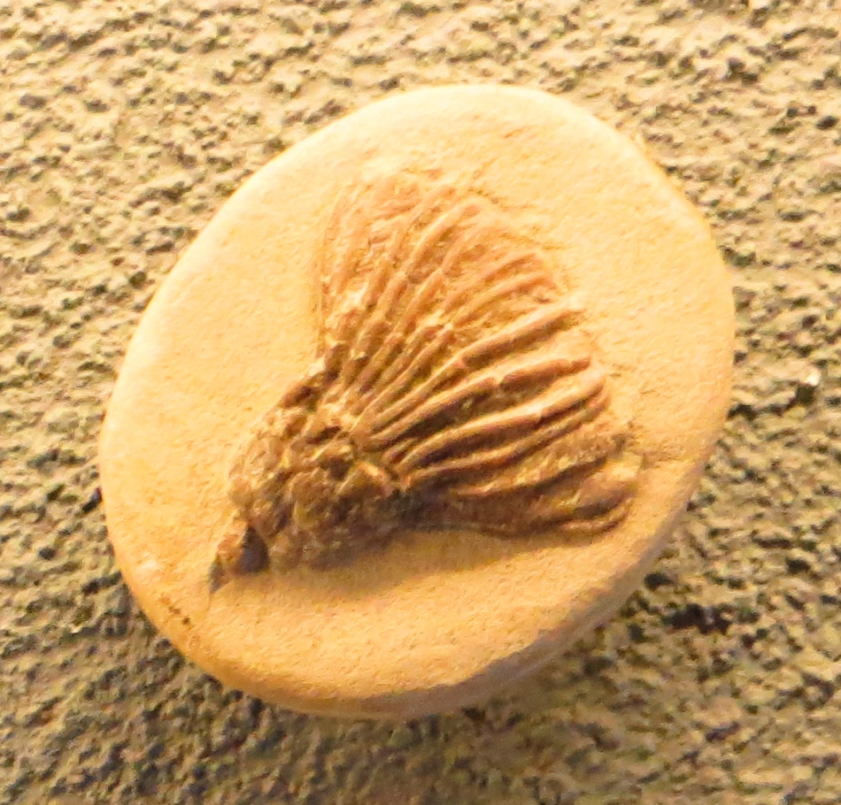
The accompanying photograph shows a Rhodocrinites kirbyi fossil displayed at Naturalis in Leiden, with its closely packed arms preserved together.[7]
The strength of these studies lies in connecting a small feature to a larger pattern. One arm records an individual's recovery. Differences among species, sizes and geological intervals allow researchers to ask why some animals were more likely to bear that record.
The survivors have a clock
A collection of regenerated arms is necessarily a collection of survivors. An animal killed outright cannot subsequently produce a healed injury. Consequently, fewer regenerated specimens could reflect fewer attacks, more effective defenses, or more lethal attacks. Gahn and Baumiller explicitly cautioned against treating regeneration frequency as a simple reading of predation intensity.[3]
There is another complication: the same injury remains visible for different lengths of time in different animals.
In a 2013 paper, the pair developed a method for estimating arm-loss rates that accounts for differences in arm length, arm number and branching. A proportion of injured individuals is a snapshot; a rate asks how frequently damage occurs. Moving between the two requires information about the body being rebuilt.[4]
Their model estimated arm loss roughly once every ten days for the living feather star Cenometra bella, compared with once every 36 days for the fossil Rhodocrinites kirbyi. These are reconstructed rates for particular examples, dependent on the method's assumptions. They should not be read as directly observed schedules or universal figures for modern and ancient seas.[4]
Living crinoids show why healing speed deserves attention. Angela Stevenson and colleagues' study, published in 2022, found that swimming feather stars regrew arms up to three times faster than nonswimmers in their comparison. Swimming ability explained differences among species better than the environmental and anatomical alternatives they tested. Within a temperate species, however, warmer conditions did accelerate regrowth.[5]
For a paleontologist, that creates a practical problem. A rapidly rebuilding animal can erase the visible stage of an injury sooner. Two assemblages with similar numbers of damaged arms need not represent the same frequency of encounters. Reading scars requires some understanding of how long they remain scars.[4][5]
Survival does not reveal the attacker's purpose
Even a convincingly predatory injury leaves an unanswered question: what was the predator trying to eat?
In a 2018 study of crinoid spines and regeneration, Valerie Syverson and colleagues distinguished the outcome of an encounter from the predator's intention. A crinoid might survive an attempted killing, or lose only part of its body during feeding. Their evidence also supported the possibility that some predators targeted organisms living on crinoids, including attached snails.[6]
This expands the scene without pretending to recover every movement. The injured animal may have been the meal, part of the meal, or the structure carrying another desirable animal. Regrowth alone cannot choose among those stories.[6]
Return to the small plates beyond the broken arm. They establish something firmer than a dramatic reconstruction: the injury was followed by growth. Its eventual death preserved evidence that an earlier injury had not been the end.
Sources
- Tomasz K. Baumiller and Forest J. Gahn, “Testing Predator-Driven Evolution with Paleozoic Crinoid Arm Regeneration.” Science 305 (2004), 1453–1455; specimen photograph in figure 1D and the eleven-assemblage comparison.
- Forest J. Gahn and Tomasz K. Baumiller, “Evolutionary History of Regeneration in Crinoids (Echinodermata).” Integrative and Comparative Biology 50 (2010), 514a–514m; recognition, repair and the deep history of regeneration.
- Forest J. Gahn and Tomasz K. Baumiller, “Arm regeneration in Mississippian crinoids: evidence of intense predation pressure in the Paleozoic?” Paleobiology 31 (2005), 151–164; Le Grand assemblage and limits of injury frequencies.
- Tomasz K. Baumiller and Forest J. Gahn, “Reconstructing predation pressure on crinoids: estimating arm-loss rates from regenerating arms.” Paleobiology 39 (2013), 40–51; converting regeneration proportions into estimated rates.
- Angela Stevenson, Tadhg C. Ó Corcora, Christopher D. G. Harley and Tomasz K. Baumiller, “Ability to Swim (Not Morphology or Environment) Explains Interspecific Differences in Crinoid Arm Regrowth.” Frontiers in Marine Science, published January 13, 2022; comparisons of living feather stars.
- Valerie J. P. Syverson, Carlton E. Brett, Forest J. Gahn and Tomasz K. Baumiller, “Spinosity, regeneration, and targeting among Paleozoic crinoids and their predators.” Paleobiology 44 (2018), 290–305; partial predation and possible targets of attacks.
- Ghedoghedo, “Rhodocrinites kirbyi Naturalis.” Museum specimen photographed at Naturalis, Leiden, August 26, 2013; Wikimedia Commons, CC BY-SA 3.0.