Fritz F.'s hand survives in wax. Made by Lotte Volger in 1929, the model records a spreading cowpox infection across its back. Yet when the University of Zurich exhibited it in August 2019, the accompanying account could not explain how he had become infected. Vaccination was one possibility; contact with an infected cow was another. The medical files had been discarded.[1]
The uncertainty is striking because the object promises such intimate knowledge. Here is the shape of an individual hand, preserved for close inspection. Its owner, however, remains partly beyond reach. How should a medical museum remember someone whose symptoms are easier to recover than his circumstances?
Zurich's Museum of Wax Moulages offers a particularly revealing place to ask. A moulage is a cast model of a patient's affected body surface. These objects began as tools for teaching and documentation; preserving them has also made them records of the people whose bodies supplied the lessons.[2][7]
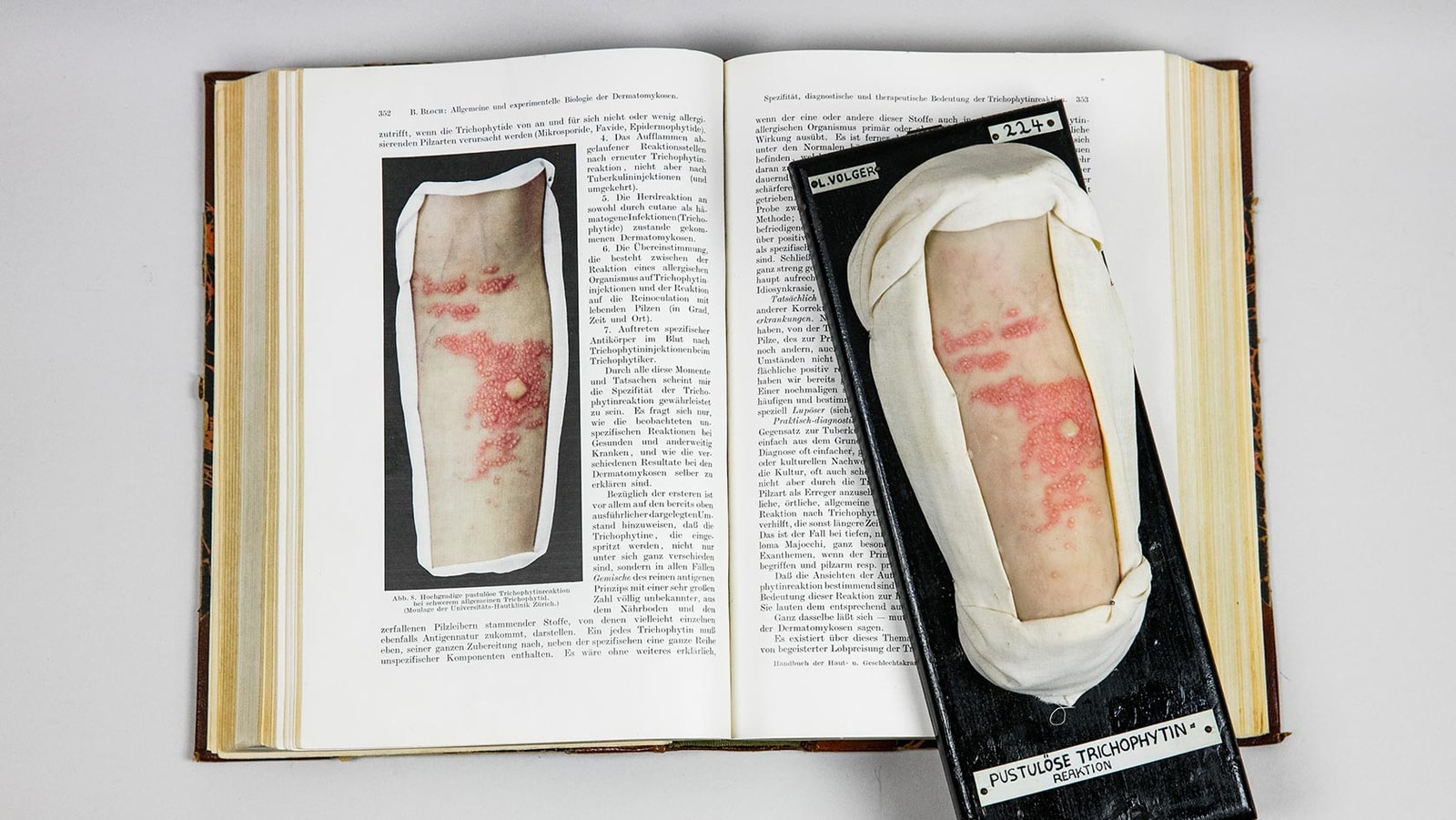
A likeness made through skilled hands
A moulage starts with an impression taken from the skin. In the Zurich technique, a negative mold receives a warm wax mixture. The maker then corrects the cast, adds details such as scales or blisters, inserts individual hairs and paints the coloration. Traditionally, the finished model rests on a black wooden board edged with white fabric, accompanied by identifying information.[2]
That sequence makes the object both an impression and an interpretation. The mold preserves surface form, while the maker's decisions help render disease visible. A visitor is looking at a patient's body through another person's trained observation. The work deserves attention as a clinical document and as skilled labor.
The preserved hand also illustrates a limit of visual realism. Even a remarkably faithful surface cannot disclose an occupation, an exposure or a treatment history by itself. The 2019 exhibition acknowledged alternative explanations for Fritz F.'s infection. Keeping those alternatives visible protects the patient from acquiring an invented biography.[1]
The collection almost disappeared
Volger brought her craft to Zurich in 1918. In the dermatology clinic completed in 1924, the models were kept beside the lecture hall, ready for teaching. Their physical presence was useful, but it demanded storage and care. Later, improved color photography and projection helped displace them.[4]
According to the museum's institutional history, Elsbeth Stoiber was instructed in 1972 to discard the dermatological collection. She refused to carry out the task and worked to preserve it. A 1979 exhibition helped renew interest; a dedicated museum room opened in 1993.[4]
This account gives preservation a human agent. Stoiber resisted treating a change in teaching technology as the end of the collection's value. The museum's own retrospective is the basis for this reconstruction.
The episode changes the meaning of the cabinets. An obsolete teaching aid can remain evidence for questions its original users never asked. What did clinicians choose to record? Whose work made a condition legible? Which details of a patient's life accompanied the image, and which were allowed to disappear?
Reconnecting an object with a life
Missing case files do not make every patient history irretrievable. For its 2005 exhibition on surgical moulages, the museum combined surviving patient information with research into published case reports. That work recovered aspects of the personal and social circumstances behind models made between 1919 and 1927. Some had documented unusual findings or attempts at new surgical treatments.[5]
Another exhibition that year examined models connected with animal experiments, self-experiments and clinical research. These uses complicate the idea of a cabinet containing only representative examples of disease. A model might belong to an experiment, a treatment sequence or an individual clinical puzzle.[5]
For remembrance, the connection matters. A diagnosis can explain why a body part attracted medical attention. A linked record can begin to explain what was happening to the person. Where that connection survives, curators can restore context. Where it fails, an honest gap is more informative than a confident story assembled from appearances.
This is also why the museum's continuing teaching role matters. Its exhibition still serves medical students while giving other visitors explanations of the objects' changing meanings.[3] The patient remains involved in education, but the lesson can now include the limits of what the preserved image permits us to know.
The label belongs to the patient record
Conservation makes this responsibility tangible. Recommendations developed through a 2008–2010 project at the German Hygiene Museum in Dresden treat a moulage as an entire object: wax, mounting, support, case and labels all require preservation. They also advise retaining evidence of earlier interventions, including amended disease descriptions.[6]
A changed diagnosis is therefore part of the history worth keeping. It can show that medical knowledge moved while the object remained. Removing an untidy label could make the display cleaner while stripping away evidence of how the patient was understood.
The same recommendations caution that restoration involves subjective judgment. Filling gaps or retouching paint can affect how the represented condition reads; such work should be exceptional and involve conservation and medical expertise.[6] Care for these objects includes knowing when an improvement might obscure evidence.
Seen this way, Zurich's collection performs a modest kind of commemoration. It holds onto traces of particular people, the makers who studied them and the changing judgments attached to their bodies. Its value depends partly on resisting the temptation to make those traces seem complete.
Fritz F.'s hand can still be studied. The uncertainty beside it deserves to survive too.
Sources
- University of Zurich Medical Collection, “ISEMPH Exhibition” (August 13, 2019), object 1 — Fritz F.'s 1929 hand moulage, possible routes of infection and the loss of medical files.
- Museum of Wax Moulages, University and University Hospital Zurich, “Moulage Production Technique” — casting, modeling, coloring and mounting.
- Museum of Wax Moulages, “Exhibition” — the collection's continuing teaching function and historical interpretation.
- Museum of Wax Moulages, “Moulage History in Zurich” — the clinic collection, Elsbeth Stoiber's rescue and the museum's establishment.
- Museum of Wax Moulages, “Side Showcase,” especially the two 2005 exhibitions — research into patient histories, surgery and experimentation.
- German Hygiene Museum Dresden project, “Recommendations for the Preservation of Wax Moulages at Universities and Hospitals and in Museums and Other Collections” (2008–2010 project), especially pages 3, 6–8 — institutional copy hosted by ICOM's university museums committee.
- University of Zurich, “Moulagenmuseum” — institutional collection overview and source of the photograph; embedded image metadata credits Lukas Linder.