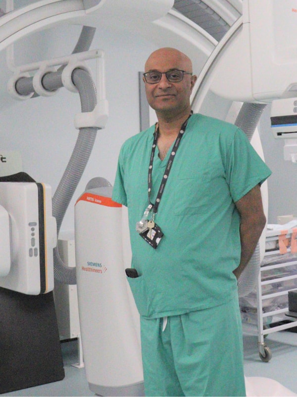
The photograph places a specialist beside the machinery that lets him navigate inside an artery. When Addenbrooke's Hospital opened its second biplane angiography suite in July 2023, the expansion included two additional interventional neuroradiology consultants and support staff. The equipment and the people were parts of the same promise: getting stroke patients to clot removal sooner.[1]
Mechanical thrombectomy has an unusually tangible objective. A clot obstructing a brain artery can be caught and withdrawn through a catheter. Yet the object removed cannot tell the whole story of the person on the table. The treatment aims to reconnect threatened brain tissue to its blood supply; recovery depends on what that tissue can still do.
A route through the blood vessels
In the procedure described by Leeds Teaching Hospitals, the operator enters through the femoral artery in the groin and advances a catheter toward the blocked brain vessel. Contrast makes the vessels visible under X-rays. A smaller catheter carries a mesh device, the stent retriever, into the clot. The mesh expands and engages the obstruction; withdrawal, accompanied by suction, aims to bring the clot out while limiting fragments escaping farther downstream.[2]
That sequence explains why a successful retrieval is an achievement in its own right. The operator must reach the obstruction, capture it, and remove it through a branching vascular route. Leeds describes repeat attempts as passes, continued until removal succeeds or further attempts become unsafe. Some clots cannot be removed. Vessel injury and bleeding are among the risks.[2]
The visible endpoint is renewed passage of blood. The biological objective lies farther along: supplying tissue whose function may still be recoverable.
The scan estimates an opportunity
The distinction between an ischemic core and a penumbra helps explain the opportunity. The core represents the most severely injured region; the penumbra is under-supplied tissue that may remain salvageable. Restoring circulation can prevent threatened tissue from progressing to infarction. But an imaging estimate of core is a prediction, with uncertainty, rather than a cell-by-cell inventory of irreversible loss.[3]
The 2026 AHA/ASA guideline makes that last point explicit: changes in CT density, diffusion imaging, or estimated cerebral blood flow predict infarction without proving that every affected part is already beyond rescue. This matters because an image can guide a treatment decision without drawing an infallible boundary around living and dead tissue.[3]
Disability is the outcome that matters
The 2016 HERMES analysis pooled individual data from five randomized trials: 1,287 patients, of whom 634 were assigned to thrombectomy and 653 to control care. Its central outcome was disability after 90 days, measured on the modified Rankin scale. Thrombectomy improved the distribution of disability outcomes, with an adjusted common odds ratio of 2.49 and a 95% confidence interval of 1.76–3.53.[4]
The investigators estimated that treating 2.6 patients produced one additional patient with at least one level less disability. That figure describes a shift on a disability scale. It should not be read as a promise that one in every few procedures produces complete recovery.[4]
This is why the endpoint matters. Removing an obstruction demonstrates a physical success. Being able to manage daily life measures a consequence that patients and families must live with.
How the later window opened
In 2018, DEFUSE 3 tested thrombectomy 6–16 hours after patients were last known well. Researchers enrolled people with specified large-artery blockages and favorable perfusion-imaging findings, including an estimated core below 70 milliliters and a ratio of hypoperfused tissue to core of at least 1.8. Those were research enrollment criteria for selecting a population with a plausible opportunity for tissue rescue.[5]
The trial randomized 182 patients and stopped early for efficacy. At 90 days, 45% of the thrombectomy group and 17% of the medical-therapy group were functionally independent, defined as a modified Rankin score of 0–2. That is a 28-percentage-point difference in this selected population. Functional independence can still include residual symptoms or disability.[5]
Subsequent evidence widened eligibility. The 2026 guideline supports thrombectomy for selected patients with larger predicted cores and selected basilar-artery occlusions in the posterior circulation. DEFUSE 3's thresholds remain a landmark research framework, rather than a universal rule for today's care.[3]
An open window still rewards speed
A separate 2016 HERMES time analysis examined patients who achieved substantial reperfusion. Among 390 such patients, each additional hour to reperfusion was associated with a 5.2-percentage-point lower probability of functional independence; the confidence interval for that reduction was 2.1–8.3 points. Treatment delays were not randomly assigned, so this association is not a precise personal countdown.[6]
It nevertheless explains why extending eligibility and reducing delay fit together. Imaging may reveal a remaining opportunity, while faster treatment helps preserve it. A later outer boundary gives a stroke team more people to assess; it gives a bystander no reason to wait.
For sudden stroke symptoms—such as one-sided weakness, speech difficulty, or new loss of balance or vision—call emergency services immediately, note when symptoms began, and use an ambulance. The CDC advises against driving to the hospital.[7]
The room in the photograph is where navigation, imaging, and clinical judgment converge. Its purpose reaches beyond the retrieved clot, toward the daily activities a person may be able to recover.
Sources
- Cambridge University Hospitals, “New bi-plane angiogram suite opens,” 7 July 2023—Addenbrooke's service expansion, staffing, and the photograph of Yogish Joshi.
- Leeds Teaching Hospitals NHS Trust, “Mechanical Thrombectomy”—catheter access, stent retrieval, suction, repeated passes, and procedural risks.
- Prabhakaran et al., AHA/ASA acute ischemic stroke guideline, January 2026—sections 4.7.2–4.7.3, especially page e54; SETRAC-hosted full text.
- Mayank Goyal and colleagues, “Endovascular thrombectomy after large-vessel ischaemic stroke: a meta-analysis of individual patient data from five randomised trials,” The Lancet, 2016—HERMES population, disability endpoint, treatment effect, and number needed to treat.
- Gregory W. Albers and colleagues, “Thrombectomy for Stroke at 6 to 16 Hours with Selection by Perfusion Imaging,” New England Journal of Medicine, 2018—DEFUSE 3 selection criteria, early stopping, and functional outcomes.
- Jeffrey L. Saver and colleagues, “Time to Treatment With Endovascular Thrombectomy and Outcomes From Ischemic Stroke: A Meta-analysis,” JAMA, 2016—association between reperfusion delay and functional independence.
- Centers for Disease Control and Prevention, “Signs and Symptoms of Stroke,” updated 19 May 2026—symptom recognition, immediate emergency contact, and ambulance transport.