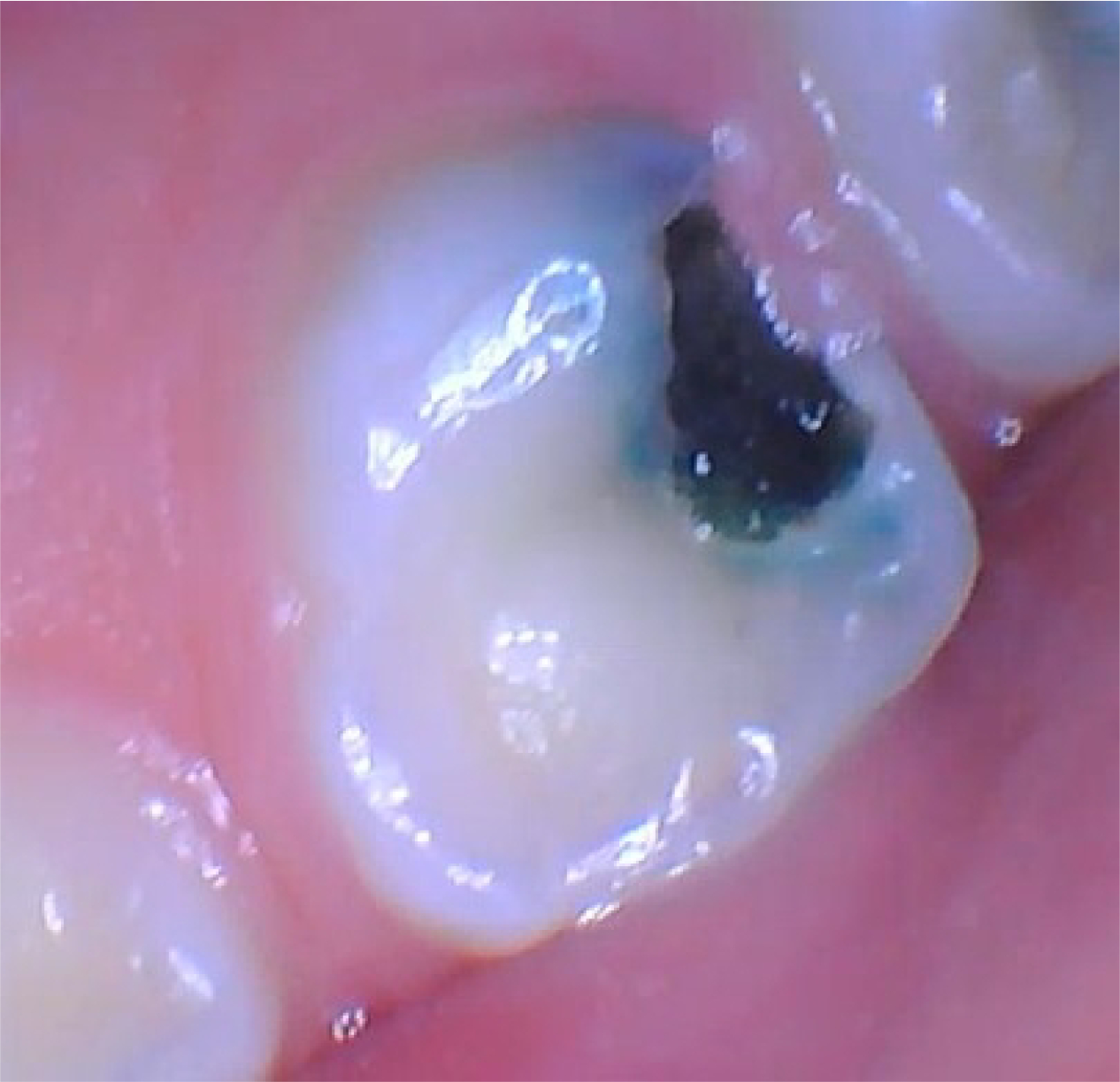
The black patch in the cover photograph looks like an endpoint. Its dark color resembles the staining patients and clinicians expect after silver diamine fluoride, or SDF, touches decayed dentin. Yet at the eight-month examination this study tooth's lesion was still soft and had grown. Color alone had not established arrest—and the trial report does not identify whether the pictured tooth received SDF or the tinted placebo.[7]
That distinction opens a history larger than one disappointing tooth. SDF had two distinct institutional arrivals. In 1969, Mizuho Nishino completed an Osaka University doctoral study on ammoniacal silver fluoride and the progression of caries in primary teeth. A product followed in Japan in 1970, and Nishino's group published an English-language article on the material in 1972.[1][2][3] More than four decades later, SDF's U.S. regulatory entry came through a door marked dentin sensitivity, before clinicians adopted it off label to arrest cavities without first drilling them.[3]
The chemistry did not cross the Pacific alone. Its meaning changed with the care system around it. Japanese records emphasize formulation, approval and early clinical applications; later U.S. guidance emphasizes reaching children and other patients for whom an ordinary restoration might be delayed, difficult or impossible. Comparing those institutional pathways explains both SDF's power and its limit: a liquid can interrupt a disease process, but it cannot make diagnosis, follow-up or lost tooth structure disappear.
Osaka, 1969: joining silver and fluoride in one liquid
Nishino's thesis record is unusually precise. Osaka University dates the degree to March 28, 1969 and describes a study of ammoniacal silver fluoride for suppressing the progression of caries in deciduous teeth.[1] In 1972, Reichi Yamaga, Nishino, S. Yoshida and I. Yokomizo published Diammine Silver Fluoride and Its Clinical Application in the university's dental journal.[2] A later evidence review traces the commercial product Saforide to Japanese approval in 1970.[3]
The innovation joined two already familiar therapeutic ideas. Silver compounds had a long antimicrobial history in dentistry; fluoride could make mineral less vulnerable to the repeated acid challenge produced when a cariogenic biofilm metabolizes sugar. Ammonia complexes and stabilizes silver(I), allowing a concentrated alkaline solution containing silver and fluoride. The 38% formulation used in current U.S. guidance is not 38% fluoride, but a concentrated SDF solution containing silver, fluoride and ammonia.[3]
This was not a filling in liquid form. A restoration replaces missing anatomy and gives a tooth a cleanable, load-bearing surface. SDF instead offered a nonrestorative intervention: apply a small amount directly to accessible decay and try to turn soft, active dentin into hard, inactive tissue. Early Japanese authors described pediatric caries arrest and prevention, secondary-caries control and desensitization, while warning that dark staining limited acceptability—particularly on anterior teeth.[3] A material that could be painted on nonetheless promised a very different encounter from excavation, preparation and placement of a restoration.
Japan's first arrival therefore began with a material question: could silver's action and fluoride's action be combined in a formulation that arrested caries? The answer became credible enough for clinical use well before every molecular step was settled. That ordering matters. Medicine often learns that an intervention works before it can specify exactly which of several plausible pathways contributes most in living tissue.
United States, 2014: the material becomes an access strategy
SDF's American entry had an odd shape. In 2014, the U.S. Food and Drug Administration cleared a 38% product for adult dentin hypersensitivity; the first commercial product became available in 2015. Caries arrest was—and in the final 2026 trial report remained—an off-label use.[3][8] The substance was old. Its place in American practice was new.
The unmet need made that second arrival consequential. Conventional restorative dentistry may require local anesthesia, rotary instruments, moisture control, cooperation for a sustained procedure and, for some very young or medically complex patients, sedation or general anesthesia. SDF needs case selection and careful isolation, but the application itself is brief and does not require removal of sound tooth tissue. The American Academy of Pediatric Dentistry's chairside guide accordingly identifies patients with multiple lesions, behavioral or medical management challenges, hard-to-treat cavities, or limited access to dental care as groups who may benefit.[4]
This widened the intervention's practical role. SDF could buy time until a child became able to tolerate definitive restoration. It could help manage a primary tooth approaching natural exfoliation. It could take a treatment into a school or community clinic where a full restorative setup was unavailable. In the New York City CariedAway trial, registered nurses and dental hygienists, working under pediatric-dentist supervision, delivered a one-time package of 38% SDF plus fluoride varnish in schools; the comparison group received glass-ionomer sealants, atraumatic restorations and the same varnish.[9]
In the trial's 2023 restricted analysis, 2,998 recruited-and-treated children were eligible for analysis and 1,398 were observed at follow-up after pandemic disruption. Among children with baseline caries, 56% in the SDF-plus-varnish group and 46% in the sealant/atraumatic-restoration-plus-varnish group had lesions classified as arrested; 81% and 82%, respectively, remained free of new caries. The package met its prespecified noninferiority criterion.[9] That supports a simpler school-delivered approach, but only within its frame: the study compared treatment bundles rather than isolating SDF, and the substantial loss to follow-up warrants caution when applying its result elsewhere.
The pathways were not mirror images. Japan's ran from university chemistry to a nationally approved product used for pediatric caries, secondary caries and sensitivity, with staining constraining anterior use. The U.S. pathway began with device clearance for adult hypersensitivity, followed by off-label pediatric adoption, professional guidance and school-based delivery. These were not two discoveries of SDF, but two institutional histories of the same material.
This Japan–U.S. comparison is necessarily selective. Decades of clinical use and trials elsewhere supplied important evidence later incorporated into American practice and global guidance. The comparison here concerns what each documentary record foregrounds—not two isolated national traditions.
What the liquid changes inside a lesion
The simplest account of SDF says that silver kills bacteria while fluoride rebuilds mineral. It is directionally useful and biologically incomplete. Caries is not a single pathogen sitting in a hole. It is a local ecological process in which biofilm metabolism repeatedly lowers pH, mineral leaves enamel and dentin, and the organic dentin matrix becomes vulnerable.[10]
Laboratory and extracted-tooth studies suggest several effects after SDF enters porous carious tissue. Silver can interfere with bacterial membranes, enzymes and replication; fluoride can reduce further mineral loss and favor mineral deposition; and SDF can inhibit matrix metalloproteinases and cysteine cathepsins that participate in collagen degradation.[10] These routes may reinforce one another, but the mechanism review found that clinical trials had tested outcomes, not isolated the contribution of each molecular pathway. “It sterilizes the cavity” is therefore too strong. “It can push an active lesion toward a harder, less actively demineralizing state” better matches the evidence.
The stain has its own uncertain chemistry. Silver-containing reaction products can darken through reduction or photochemical change, potentially producing metallic silver and silver-chloride-derived particles among other species. A 2024 critical review cautions that the dominant reaction in the mouth has not been pinned to one universal black compound.[11] When darkening follows confirmed SDF application, silver chemistry is one explanation; black color itself is neither specific to SDF nor proof of arrest, as some trial lesions were black before treatment.[7][8] It does not, by itself, measure lesion hardness, bacterial activity or future progression.
That is why the cover image is not a paradox. The pathways to blackening and arrest overlap, but they are not identical. Lesion depth, plaque exposure, disease environment and treatment exposure influence arrest probability across studies, but the photograph cannot establish why this particular lesion remained active—or even which study liquid it received.
Evidence learned to separate color from outcome
American guidance initially moved with appropriate caution. The AAPD's 2017 clinical practice guideline made a conditional recommendation for 38% SDF in cavitated primary-tooth lesions as part of comprehensive caries management; the underlying evidence was rated low quality, and dark staining made examiner blinding difficult.[3] The current chairside guide operationalizes the follow-up rule: treated lesions usually become black and hard, a check at roughly two to four weeks is advisable, and reapplication should depend on color, hardness and evidence of progression rather than color alone.[4]
A 2024 Cochrane review likewise judged the evidence for arrest in primary teeth to be low certainty and the evidence in permanent teeth very uncertain. Its search ended in June 2023, so it could not include the final 2026 phase III report.[12] The chronology matters: a later positive trial can strengthen an evidence base without retroactively changing what an earlier systematic review was able to assess.
The phase III U.S. trial gives that distinction numerical force. Its final report, published in July 2026, enrolled 830 children aged 12 to 71 months with severe early childhood caries and began with 1,987 active cavitated lesions. At six months after one application, 54.0% of SDF-treated lesions versus 22.5% of placebo-treated lesions were arrested—a 31.5-percentage-point difference (99.9% confidence interval, 21.5 to 41.6).[8] Of the 830 enrolled children, 584, or 70%, completed follow-up. That is a clinically important effect, not a universal success; about half of the SDF lesions in this unusually high-risk population were still not classified as arrested.
Investigators judged arrest by a change from soft to hard dentin and calibrated color separately. The design choice was necessary: although every lesion was active at baseline, 41, or 2.1%, were already black.[8] An interim report from the same trial supplied the cover photograph as an even plainer warning: a black lesion documented during the trial that remained soft and enlarged.[7] The eye can notice a color. It cannot complete the examination.
Guidance also became more specific as the evidence base matured. In March 2026, the World Health Organization issued a strong recommendation, based on moderate-certainty evidence, for 38% SDF applied twice yearly to cavitated lesions in primary teeth and to cavitated root-surface lesions in permanent teeth, provided there are no signs or symptoms of pulp involvement.[5][6] That wording is deliberately bounded: it is not a blanket recommendation for every coronal cavity in a permanent tooth.
The boundary neither arrival removed
SDF is most useful when the lesion is accessible to the applicator and the pulp is not already declaring a deeper problem. Spontaneous pain, pulp exposure, clinical or radiographic signs of pulpal inflammation or necrosis, swelling, a fistula or periapical disease rule out SDF-only management and require diagnostic and definitive dental evaluation. Radiographs should be obtained when feasible to assess lesion depth. WHO and AAPD guidance also address relevant product allergies, protection of surrounding soft tissue, informed consent about persistent black dental staining and the need for reassessment.[4][6]
Nor does arrest restore form or mechanical strength. A treated tooth may still need glass ionomer, composite, a crown, pulp therapy or extraction. SDF may be an interim step, a repeated maintenance treatment or one part of a later restoration, and it should sit within comprehensive caries care that includes plaque control, fluoride toothpaste, dietary management and risk-based reassessment.[4][6] The choice depends on the tooth, the person, the care setting and what follow-up can actually be delivered.
This is where the comparison between Osaka and the American school clinic becomes most revealing. Japan's early development made a concentrated liquid clinically usable; later U.S. institutions placed that liquid inside an access pathway. Neither history turned the black stain into a diagnosis. The enduring achievement is more modest and more valuable: suitable active cavitated lesions that once seemed to demand an immediate drill can sometimes be interrupted with a brush—provided a trained dental clinician reassesses hardness, symptoms and progression, then decides what the tooth still needs.
Sources
- Mizuho Nishino, “Studies on the Topical Application of Ammoniacal Silver Fluoride for the Arrest of Dental Caries” (doctoral thesis record, Osaka University, degree awarded March 28, 1969) — institutional record for the Japanese research origin and date.
- Reichi Yamaga, Mizuho Nishino, S. Yoshida and I. Yokomizo, “Diammine Silver Fluoride and Its Clinical Application,” Journal of Osaka University Dental School 12 (1972), 1–20 — bibliographic record for the early clinical publication.
- Yasmi O. Crystal et al., “Evidence-Based Dentistry Update on Silver Diamine Fluoride,” Dental Clinics of North America 63(1), 2019 — history of the 1970 Japanese approval, 2014–2015 U.S. entry, formulation, adoption and evidence context.
- American Academy of Pediatric Dentistry, “Chairside Guide: Silver Diamine Fluoride in the Management of Dental Caries Lesions” — current case-selection, consent, application, two-to-four-week follow-up and reapplication guidance.
- World Health Organization, WHO Guideline on Environmentally Friendly and Less Invasive Oral Health Care for Preventing and Managing Dental Caries (March 5, 2026) — current recommendation, evidence grade, lesion boundaries and implementation considerations.
- World Health Organization, “Recommendation 5: Silver Diamine Fluoride,” in the 2026 WHO caries guideline — exact twice-yearly regimen, eligible lesions, pulp-status boundary and certainty assessment.
- Margherita Fontana et al., “A Randomized Clinical Trial to Assess Caries Arrest by Using Silver Diamine Fluoride in U.S. Children—Interim Findings,” Pediatric Dentistry 46(1), 2024 — figure 2 is the clinical photograph used as the cover image and documents a black but still-active study lesion without identifying treatment assignment.
- Margherita Fontana et al., “Efficacy of Silver Diamine Fluoride on Young Children With Severe Early Childhood Caries,” JAMA Pediatrics (published online July 27, 2026) — final phase III trial population, completion, hardness-based outcome and arrest estimates.
- Ryan Richard Ruff, Tamarinda Barry-Godín and Richard Niederman, “Effect of Silver Diamine Fluoride on Caries Arrest and Prevention: The CariedAway School-Based Randomized Clinical Trial,” JAMA Network Open 6(2), 2023 — school delivery, treatment bundles, outcomes and follow-up limitations.
- Irene Shuping Zhao et al., “Mechanisms of Silver Diamine Fluoride on Arresting Caries: A Literature Review,” International Dental Journal 68(2), 2018 — laboratory evidence on antimicrobial, mineral and anti-protease pathways and the limits of mechanistic inference.
- Martin J. Thomas et al., “Physicochemical Properties, Biological Chemistry and Mechanisms of Action of Caries-Arresting Diammine-Silver(I) Fluoride and Silver(I)-Fluoride Solutions for Clinical Use: A Critical Review,” Frontiers in Oral Health 5, 2024 — uncertainty around the silver species and reactions responsible for dark staining in vivo.
- Anne-Marie Glenny et al., “Silver Diamine Fluoride for Managing Caries in Children and Adults,” Cochrane Database of Systematic Reviews 2024(7) — evidence-certainty assessment and June 2023 search date.