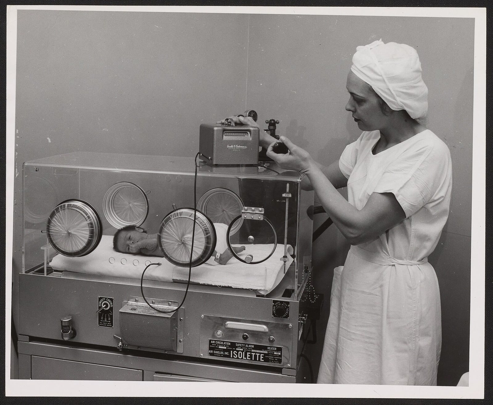
The nurse has one hand on a valve and her eyes on a meter. Below her, a premature infant lies inside a glass incubator, watched through round access ports. The photograph was made around 1959, after a decade in which neonatal medicine discovered that the gas keeping a baby alive could also take the baby's sight.[9]
The meter is the important object. Earlier incubators could be filled with oxygen-rich air for days or weeks, as if oxygen were simply a better atmosphere for a struggling infant. By the end of the 1950s, the nursery had learned to treat it as an exposure: something with a concentration, a duration, a benefit, and a toxicity.
That lesson is often compressed into a clean triumph—doctors found that too much oxygen caused blindness, so they turned it down. The actual reconstruction is harder. A mysterious eye disease appeared among the premature infants whom new nurseries were becoming best equipped to save. Small trials implicated oxygen, but withholding a breathing treatment looked dangerous. A national study appeared to show that restriction protected sight without reducing survival. Decades later, much larger trials demonstrated the boundary that the first victory had obscured: lower oxygen targets can reduce severe retinopathy while increasing death.[6][7]
The achievement was therefore not finding one safe number. It was learning that there would never be a morally simple side of the dial.
1942: a new disease appears behind the lens
In 1942, Boston ophthalmologist Theodore Terry described an unusual disorder in premature infants. He called its late appearance retrolental fibroplasia: fibrous tissue forming behind the lens. By the late 1940s, nurseries in the United States were seeing enough children with detached retinas and profound visual loss for clinicians to speak of an epidemic.[1][2]
The name described the scar, not the beginning of the injury. Modern accounts call the disease retinopathy of prematurity, or ROP, and place its origin in a retina whose blood vessels have not finished growing. High oxygen exposure early after preterm birth can suppress normal vascular development. When the developing retina later demands more oxygen than its incomplete circulation can deliver, the resulting relative hypoxia can drive disordered new vessels, bleeding, fibrosis, and detachment. This two-phase model was assembled later; the physicians facing the first wave saw only the delayed wreckage.[1]
That delay made cause difficult to recognize. Prematurity itself was an obvious common factor, and investigators pursued infection, nutrition, light, vitamin deficiencies, blood transfusion, and many other candidates. Oxygen was especially difficult to indict because the sickest infants were both the most likely to receive it and the most likely to develop complications. Association could reflect treatment, illness severity, or both.[1][2]
There was also an emotional asymmetry. Cyanosis and respiratory distress were visible now. Retinal injury emerged later, often after the breathing crisis had passed. Turning oxygen up looked like rescue; leaving it low looked like neglect.
1951–1952: the geographic clue becomes a nursery trial
The pattern sharpened across hospitals and countries. On July 14, 1951, Australian pediatrician Kate Campbell reported retrolental fibroplasia in 23 of 123 premature infants managed with intensive oxygen, compared with 3 of 44 who received more moderate therapy. American and British nurseries also seemed to diverge: the disease was common where oxygen had become abundant and much rarer where the gas was used sparingly.[2]
Those comparisons were powerful clues, but not verdicts. Oxygen practice varied alongside equipment, nursing, survival, referral, and the maturity of babies admitted. A nursery that kept smaller infants alive long enough to develop ROP could look worse precisely because its immediate care was better.
At Gallinger Municipal Hospital in Washington, ophthalmology resident Arnall Patz and pediatrician Leroy Hoeck tried to separate the exposure from that background. Their 1952 controlled study assigned premature infants to prolonged high oxygen—roughly 65% to 70% for four to seven weeks—or to concentrations below 40%, given for shorter periods according to clinical need. Among 28 infants in the high-oxygen arm, investigators recorded 10 with early retinal changes and 7 with severe disease. Among 37 infants in the lower-oxygen arm, 6 had early changes and none had severe disease.[3]
The contrast was startling. The design was not modern: assignment alternated rather than using concealed randomization, the sample was small, and the published report did not provide the survival evidence needed to calm every pediatrician. Patz and his colleagues themselves asked for more rigidly controlled observations.[3] Their result made unrestricted oxygen look dangerous; it did not yet make restriction look safe.
1953–1954: telegrams turn suspicion into a national experiment
The next study was built around that ethical tension. Eighteen hospitals joined a cooperative trial that ran from July 1, 1953, through June 30, 1954. Eligible infants weighed 1,500 grams or less and had survived their first 48 hours. Each nursery telegraphed an enrollment to the coordinating center at Detroit's Kresge Eye Institute; the assignment came back by telegram.[4][5]
During the first three months, one infant in each set of three was assigned to routine oxygen and two to curtailed oxygen. “Routine” meant concentrations above 50% for 28 days. In the curtailed arm, oxygen was supplied when the pediatrician believed the infant needed it and was not to exceed 50%. The protocol made mortality the first gate: if survival did not differ after three months, later infants would all enter the curtailed regimen, reducing the number exposed to the suspected cause.[4][5]
That planned pivot occurred. In the concurrent phase, 15 of 68 infants assigned routine oxygen died, compared with 36 of 144 assigned curtailed oxygen—22% versus 25%, a difference the investigators did not consider statistically significant. The remaining enrollment therefore used curtailed oxygen.[5]
Across the full year, 786 infants entered the study. Of them, 166 died before 40 days, and 34 survivors lacked adequate eye follow-up, leaving 586 for the main relationship between oxygen and retinal disease. Among singleton survivors with adequate examinations, scarring ROP appeared in 17% of the routine-oxygen group and 5% of the curtailed group. Early retinal vessel changes were also much more common after routine exposure.[4][5]
On September 19, 1954, V. Everett Kinsey presented the preliminary findings in New York. An American Academy of Pediatrics editorial followed that November, and the final 63-page report appeared in October 1956. Duration mattered strongly: the longer the exposure, especially during the first days and weeks, the greater the risk. Restriction spread, and the first epidemic of oxygen-associated childhood blindness fell sharply.[2][4]
The study answered its immediate question. It also contained limits that later retellings can erase. It enrolled only babies who survived 48 hours, tested broad treatment policies rather than a continuously measured blood-oxygen target, and compared mortality concurrently for only the first part of the year. “No significant difference” in that sample was not proof that less oxygen was harmless for every premature infant.
The 1959 photograph shows the new problem correctly
The Beckman meter in the cover photograph sits on top of the incubator, connected to the chamber while the nurse adjusts a valve. Its catalogue describes the scene as an effort to provide “just the right oxygen mixture.”[9] That phrase sounds ordinary now. In 1959, it marked a new way of thinking.
The machine measured oxygen concentration in the incubator—the fraction of the surrounding gas that was oxygen. It did not continuously report how much oxygen was reaching this particular infant's blood and tissues. Those are related quantities, not interchangeable ones. Lung disease, circulation, ventilation, hemoglobin, apnea, and the fit of respiratory support all stand between gas delivered and oxygen achieved.
Later blood-gas testing and pulse oximetry moved the feedback loop closer to the baby. Clinicians could titrate support against an infant's oxygen saturation rather than rely only on a chamber concentration and visible cyanosis. But a better meter did not abolish the tradeoff. It made narrower comparisons possible.[1][7]
1960–1973: the victory acquires a mortality shadow
A later empirical warning came from hospital records rather than a new randomized trial. In 1960, Mary Ellen Avery and Ella Oppenheimer compared two eras at Johns Hopkins: 1944–1948, when oxygen had been used liberally, and 1954–1958, after restriction. Among infants weighing 1.0 to 2.5 kilograms, deaths between 30 minutes and six days after birth rose from 95 of 1,152 (8%) to 186 of 1,492 (13%). Deaths attributed to hyaline membrane disease rose from 17 to 56.[10]
The change was alarming, not conclusive. A before-and-after hospital comparison cannot isolate oxygen policy from changes in case mix, diagnosis, resuscitation, infection, drugs, or other nursery practices. In 1973, statistician K. W. Cross used British population trends to produce the era's most memorable estimate: roughly 16 additional deaths for each case of blindness prevented by oxygen restriction.[11] That figure was an ecological calculation, not a count of individually linked deaths and not a randomized causal effect.
Neither analysis acquitted unrestricted oxygen; the retinal evidence against prolonged high exposure was overwhelming. They challenged the next inference—that a blanket ceiling must therefore be safe. The cooperative trial could not observe deaths before its 48-hour enrollment point, and a chamber concentration still could not reveal exactly what reached an infant's tissues. The unresolved problem was no longer whether to turn oxygen on or off. It was how to titrate between injuries.
2005–2018: the old dilemma returns inside five percentage points
The SUPPORT trial, conducted from 2005 through 2009, randomized 1,316 infants born at 24 through 27 completed weeks to oxygen-saturation targets of 85%–89% or 91%–95%. This was not a replay of the 1950s contrast between prolonged oxygen-rich incubators and use only when thought necessary. Both groups received modern intensive care, masked oximeters, and tightly separated target ranges.[6]
The result nevertheless reproduced the old tension. Among survivors, severe ROP occurred in 8.6% of the lower-target group and 17.9% of the higher-target group. But death before discharge occurred in 19.9% versus 16.2%, respectively. The combined outcome of severe ROP or death did not differ significantly. Lower looked better if the endpoint was the eye; higher looked better if the endpoint was survival.[6]
One trial can mislead, especially when the absolute difference between targets is small and bedside readings do not remain perfectly inside assigned bands. In 2018, the NeOProM Collaboration pooled individual data from 4,965 infants in five coordinated randomized trials. The primary outcome—death or major disability by 18 to 24 months—was 53.5% with the lower target and 51.6% with the higher target, not a statistically significant difference. Yet the components pulled apart: death was 19.9% versus 17.1%, while treatment for ROP was 10.9% versus 14.9%.[7]
Those numbers are not bedside instructions. They apply to extremely preterm infants studied under particular protocols, and they do not turn one range into a universal setting for every baby, phase of illness, device, or neonatal unit. They do establish the historical point with unusual clarity: reducing an exposure-related injury does not automatically improve the total outcome.
What the nursery actually learned
Retinopathy of prematurity did not disappear when routine high oxygen did. More immature infants survived, ROP occurred even without the old exposure pattern, and screening and treatment became part of neonatal care. The National Eye Institute now describes a disease with no early signs a parent can see: at-risk babies require scheduled dilated retinal examinations so progression can be found before detachment makes the damage obvious.[8]
The durable response is therefore a system, not a slogan. Oxygen is prescribed and titrated; concentration and saturation are monitored; alarms and trends matter; retinal examinations catch a delayed injury; and trials evaluate death, lung disease, bowel injury, disability, and vision together. Each part corrects a blind spot left by the others.
The 1959 photograph deserves one final look. The infant is not an illustration of oxygen toxicity, and the nurse is not staging a morality play about excess. She is doing something more consequential: measuring a treatment whose benefit cannot excuse an unknown dose. The history began when medicine learned that oxygen could injure the babies it helped save. It matured when medicine stopped pretending that the opposite direction on the dial was automatically safe.
Sources
- Sarah H. Rodriguez et al., “Retinopathy of Prematurity in the 21st Century and the Complex Impact of Supplemental Oxygen,” Journal of Clinical Medicine 12 (2023) — modern review of the first ROP epidemic, the developing retinal circulation, oxygen-associated injury, and the two-phase disease model.
- Elizabeth A. Reedy, “The Discovery of Retrolental Fibroplasia and the Role of Oxygen: A Historical Review, 1942–1956,” Neonatal Network 23 (2004) — historical reconstruction of the epidemic, competing hypotheses, and rapid practice change.
- Arnall Patz, Leroy E. Hoeck, and Edgar de la Cruz, “Studies on the Effect of High Oxygen Administration in Retrolental Fibroplasia: I. Nursery Observations,” American Journal of Ophthalmology 35 (1952) — PubMed record for the early controlled nursery study.
- V. Everett Kinsey, June Twomey Jacobus, and F. M. Hemphill, “Retrolental Fibroplasia: Cooperative Study of Retrolental Fibroplasia and the Use of Oxygen,” AMA Archives of Ophthalmology 56 (1956) — PubMed record for the final 18-hospital cooperative-study report.
- William A. Silverman, Retrolental Fibroplasia: A Modern Parable, chapter 6, “The National Cooperative Study” — detailed reconstruction of the protocol, telegram allocation, mortality gate, trial counts, contemporary doubts, and final ROP table.
- SUPPORT Study Group, “Target Ranges of Oxygen Saturation in Extremely Preterm Infants,” New England Journal of Medicine 362 (2010) — randomized comparison of 85%–89% and 91%–95% saturation targets in 1,316 infants.
- Lisa M. Askie et al., “Association Between Oxygen Saturation Targeting and Death or Disability in Extremely Preterm Infants,” JAMA 319 (2018) — full-text record of the prospectively planned individual-participant meta-analysis of 4,965 infants in five trials.
- National Eye Institute, “Retinopathy of Prematurity” (updated August 6, 2025) — current institutional overview of disease stages, risk, dilated-eye screening, and treatment.
- Science History Institute via Wikimedia Commons, “Beckman Model D Oxygen Meter in use with an infant's incubator,” Kassler Studios, circa 1959 — catalogue and download record for the archival photograph used as the article image.
- Mary Ellen Avery and Ella H. Oppenheimer, “Recent Increase in Mortality from Hyaline Membrane Disease,” Journal of Pediatrics 57 (1960) — PubMed record for the retrospective Johns Hopkins comparison before and after oxygen restriction.
- K. W. Cross, “Cost of Preventing Retrolental Fibroplasia?” The Lancet 302 (1973) — PubMed record for the ecological estimate of mortality associated with the restriction era.