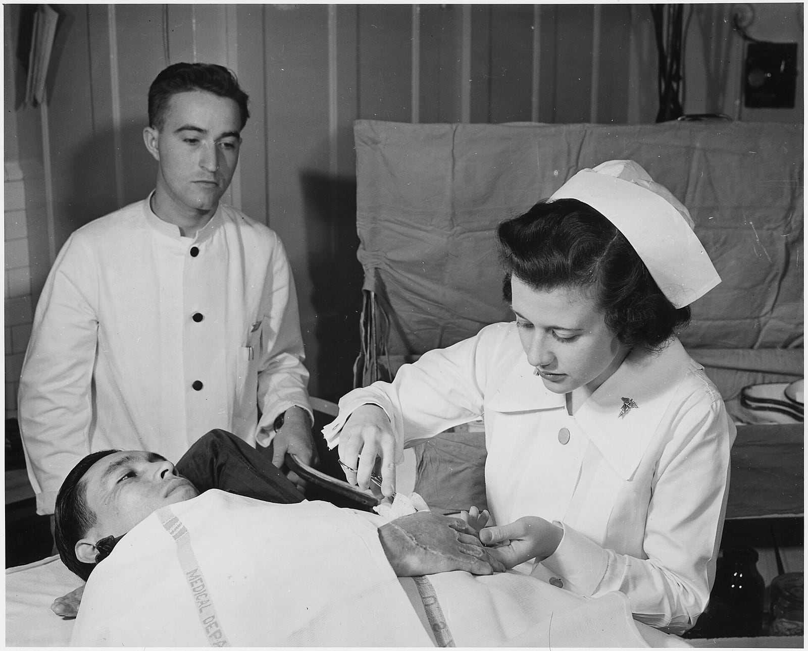
On May 23, 1943, Second Lieutenant Frances Bullock sat beside an Army hospital bed and dressed a wounded soldier's hand. The photograph makes the dressing look like the end of the story: clean gauze placed between injured skin and the world.[9] It cannot show the more difficult question underneath. What kind of environment should that covering create?
For much of the twentieth century, a dry crust looked like proof that repair was under way. The logic was visual and intuitive. Air dries the surface; the surface hardens; the wound appears sealed. Yet skin does not close a superficial wound by turning its exposed tissue into a lid. New epidermal cells have to migrate across the defect. A dehydrated scab can make that route longer and less hospitable.[1][2]
That is the evidence-backed correction: for many wounds, controlled moisture supports surface repair better than deliberate air-drying.[3][4] But the correction is often flattened into a second myth—that every wound should be made wetter, sealed more tightly, or covered with a more elaborate product. Moist wound healing is not a bath and not a brand. It is an attempt to keep the wound bed hydrated while controlling excess fluid, protecting the surrounding skin, and addressing infection, blood supply, pressure, and the cause of the injury.[5][6]
Myth: a hard scab is the repair itself
A scab is useful as a physical barrier, but its visibility gives it too much credit. In a superficial skin wound, re-epithelialization begins as keratinocytes move inward from the edges and from surviving skin structures such as hair follicles and sweat-gland ducts. They are rebuilding a surface, not pushing the old one back into place.[1]
When the exposed wound dries, its upper tissue dehydrates into a fibrous crust. Migrating epidermal cells then travel beneath that dry layer, where viable moisture remains. The scab can therefore be both evidence that the surface has dried and an obstacle the new surface must negotiate. “Dry” and “closed” are not synonyms.[1][2]
The distinction is easiest to miss when judging only by appearance. A crust can make a wound look tidier while the cellular work proceeds out of sight. Conversely, a protected wound surface may look less decisively finished even though epithelial cells have a more direct path. The old myth confused a reassuring exterior with the mechanism of repair.
1962: George Winter changed the comparison
George D. Winter's short Nature paper, published on January 20, 1962, turned that intuition into a test. Working with standardized superficial wounds in young pigs, he compared normal scab formation with conditions that prevented the surface from drying. His conclusion was narrow but disruptive: the dry scab retarded epithelialization, while preventing its formation markedly increased the rate at which new epidermis covered the wound.[1]
A 1963 follow-up by Winter and John Scales supplied the anatomical explanation. Under dry conditions, epidermis had to pass beneath dehydrated fibrous tissue. Beneath an occlusive film that kept the surface moist, it could migrate over the dermis instead. The authors reported that migration by the moist route was twice as rapid.[2]
This was animal evidence, and the boundary matters. Pig skin can model aspects of human skin repair, but it does not turn every clinical wound into the same experiment. Later in 1963, C. D. Hinman and Howard Maibach published a separate study of air exposure and occlusion in experimental human skin wounds, extending the basic comparison into people.[3] The accumulating lesson was not that scabs never protect or that occlusion solves every wound. It was that air exposure had lost its status as an automatically therapeutic act.
The 1943 photograph of Bullock predates Winter's paper by nearly nineteen years. It documents skilled wound dressing, not the moisture conditions under the gauze. That makes it a useful historical image precisely because it resists a simple before-and-after story. Clinicians had long covered wounds. The later conceptual change concerned what the cover was supposed to regulate.
What “moist” does—and what it does not mean
The word moist describes a range, not a maximum. A useful dressing may retain fluid over a dry surface, absorb fluid from a heavily draining wound, or do both at different stages. The goal is to preserve a viable wound bed without leaving the surrounding intact skin saturated.[4]
That balance changes the route available to cells. A hydrated surface supports re-epithelialization; it can also preserve a local environment in which growth signals, matrix remodeling, and new blood-vessel formation proceed. A 2013 review of moist, wet, and dry healing describes less dehydration and faster re-epithelialization under controlled moist conditions, while also stressing that wound treatment requires monitoring rather than a one-time choice of covering.[4]
The same mechanism explains why wet is not simply better than moist. Fluid that overwhelms a dressing can spread onto the skin around the wound and cause maceration—the pale, softened state produced by prolonged exposure to moisture, which can progress to tissue damage. A dressing is therefore closer to a regulator than a cap. It has to match how much fluid the wound produces and change as that production changes.[6]
Moisture is only one variable. The Agency for Healthcare Research and Quality's overview of chronic wounds places it alongside adequate blood flow, nutrition, infection control, removal of dead tissue, and management of conditions such as diabetes or vascular disease.[5] The Wound Healing Society's pressure-ulcer guideline likewise puts tissue perfusion and whole-patient assessment before aggressive moist healing, then recommends moisture balance for nonischemic wounds.[6] If circulation is inadequate, pressure continues, infection is advancing, or necrotic tissue remains, adding a moisture-retaining product does not repair the missing part of the system.
The principle is stronger than the product evidence
Once moist healing became accepted, dressings multiplied: films, foams, hydrogels, hydrocolloids, alginates, hydrofibers, and non-adherent contact layers. Their material differences are real. Some donate moisture; some hold it; some move exudate into a secondary layer. But a sound physiological principle does not prove that every product carrying its language heals every wound faster.[5]
A Cochrane review of hydrogel dressings for pressure ulcers makes that evidence boundary unusually clear. It included 11 small studies and, in its formal included-study count, 523 participants. The trials were too limited and poorly reported to determine whether hydrogels healed pressure ulcers faster or slower than alternative dressings or topical treatments.[7] That result does not restore the dry-wound myth. It separates two questions that marketing often merges: whether moisture balance matters, and whether one particular way of achieving it outperforms the others.
The separation is clinically important. A low-drainage abrasion, a surgical wound healing from its base, a venous leg ulcer, a pressure injury, an ischemic toe, and an infected bite do not present the same fluid, blood-flow, contamination, depth, or tissue problems. “Keep it moist” is a design principle. Dressing selection is an assessment problem.
For a minor cut, the modern advice is deliberately plain
The American Academy of Dermatology's guidance for a minor cut translates the principle without turning it into product theater: wash gently, stop the bleeding with pressure, apply petroleum jelly to prevent the surface from drying, cover it with a sterile bandage, and change the bandage daily. The same page advises against routine topical antibiotic ointment because it can irritate skin.[8]
That advice has a declared boundary. Persistent bleeding, a larger or deeper wound, uncertain tetanus protection after an injury from a dirty or rusty object, or increasing redness, swelling, pain, or pus changes the problem from routine home care to clinical assessment.[8] Chronic wounds and wounds complicated by poor circulation, pressure, altered immune function, or infection require cause-specific care; a moist surface cannot substitute for it.[5][6]
The best correction to the old saying is therefore not “never let a wound dry.” It is more exact: do not treat drying as if it were healing. Winter's experiment made the scab visible as an environment, not just an outcome. The decades since have made the dressing visible in the same way. Its job is not merely to hide an injury or hold medicine against it. Its job is to manage the small climate in which repair has to move.
Return to Bullock's hands in the 1943 photograph. The gauze still matters, but it no longer reads as a simple wall. It is the boundary between too little moisture and too much, between protection and inspection, between a wound's appearance and its actual progress. The enduring idea is not that covered wounds heal by magic. It is that cells repair tissue under conditions—and dryness is one of those conditions, not a cure.
Sources
- George D. Winter, “Formation of the Scab and the Rate of Epithelization of Superficial Wounds in the Skin of the Young Domestic Pig,” Nature 193, 1962 — original comparison of scab formation and protected superficial wounds.
- George D. Winter and John T. Scales, “Effect of Air Drying and Dressings on the Surface of a Wound,” Nature 197, 1963 — dehydration, the route of epidermal migration, and the reported twofold rate difference.
- C. D. Hinman and Howard Maibach, “Effect of Air Exposure and Occlusion on Experimental Human Skin Wounds,” Nature 200, 1963 — PubMed record for the early human experimental study.
- Johan P. E. Junker et al., “Clinical Impact Upon Wound Healing and Inflammation in Moist, Wet, and Dry Environments,” Advances in Wound Care 2(7), 2013 — review of re-epithelialization, inflammation, hydration, and the need for controlled monitoring.
- Agency for Healthcare Research and Quality, “Skin Substitutes for Treating Chronic Wounds” — chronic-wound assessment, moisture balance, exudate handling, circulation, infection control, and dressing functions.
- Lisa J. Gould et al., “WHS Guidelines for the Treatment of Pressure Ulcers—2023 update,” Wound Repair and Regeneration 32(1), 2024 — whole-patient assessment, perfusion, exudate control, maceration, and moisture balance for nonischemic wounds.
- Cochrane, “Hydrogel dressings for treating pressure ulcers,” 2015 — review of 11 small studies and the uncertainty around comparative healing performance.
- American Academy of Dermatology, “How to treat minor cuts,” updated February 11, 2022 — cleaning, petroleum jelly, bandaging, topical-antibiotic caution, and escalation boundaries for minor wounds.
- U.S. National Archives and Records Administration, “2nd Lieutenant Frances Bullock applies a dressing to a wounded soldier's hand in an Army hospital,” May 23, 1943 — Wikimedia Commons source page for the archival photograph.