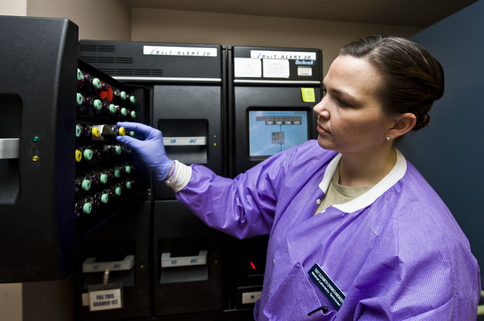
A blood-culture bottle begins with a difficult bargain. It must give a small number of living bacteria or fungi enough time and food to become detectable. At the same time, it must not let organisms carried in on the needle, bottle top, or collector's hands masquerade as an infection inside the patient.
The instrument in the cover photograph cannot settle that bargain by itself. It can incubate bottles and flag microbial growth. It cannot know whether the microbe entered the bottle from the bloodstream or from the skin during collection. That distinction is made by the entire chain: why cultures were ordered, how much blood entered each bottle, whether the draw preceded antimicrobial treatment, where separate sets were collected, which organism grew, and how the result fits the patient's condition.[1][5][6][7]
This is why a blood culture is neither a snapshot nor a generic “germ test.” It is a probability experiment followed by an identification workflow and a clinical interpretation. Most avoidable failures happen before the laboratory instrument starts watching.
The sample is the first amplifier
The culture bottle cannot grow a microbe that never entered it. In adult bloodstream infection, the organisms of interest may be unevenly distributed and sparse enough that a small draw simply misses them. More appropriate blood volume creates more opportunities to capture at least one viable organism; a second properly collected set creates another opportunity and, later, a comparison.
The U.S. Centers for Disease Control and Prevention made the adult collection target unusually concrete in guidance dated March 31, 2026. An adult set should contain 20–30 millilitres of blood, generally divided among an aerobic and an anaerobic bottle. The CDC advises collecting two to four sets for evaluation of a suspected bloodstream infection, with at least two sets providing a usual total of 40–60 millilitres. In the common two-bottle arrangement, the target is 10 millilitres per bottle.[1]
Those are adult system targets, not a universal prescription for every body. Paediatric collection uses smaller volumes adjusted to the child and the bottle system. The deeper principle is the same: the laboratory needs enough properly collected specimen for the biology it is trying to detect, without treating “more” as an unbounded good.[1][5]
Multiple sets do two jobs. They increase detection opportunity, and they create replication. If the same plausible pathogen appears across separately drawn sets, the result has a different evidential shape from a common skin organism appearing in only one bottle from one draw. Separate venipuncture sites therefore add information that one oversized specimen cannot fully replace.[1][6]
Antibiotics change the race, but the culture must not delay rescue
Once an effective antimicrobial reaches the blood, susceptible organisms may stop multiplying or die before they are sampled. Some modern culture bottles contain resins intended to adsorb antimicrobial compounds, but those materials cannot reliably undo every exposure. “Before antibiotics” is therefore a diagnostic advantage, not a ceremonial ordering rule.[3][5]
A 2019 prospective diagnostic study showed the size of that advantage within the same acute-care sequence. Investigators at seven North American emergency departments enrolled 325 adults with severe manifestations of sepsis and drew cultures before treatment and again after antimicrobial therapy began. The later draw occurred at a median of 70 minutes. Before treatment, cultures recovered at least one pathogen in 31.4% of patients; after treatment, the proportion was 19.4%. The absolute difference was 12.0 percentage points (95% confidence interval, 5.4–18.6), and the post-treatment culture's sensitivity relative to the pretreatment result was 52.9%.[3]
That study does not justify holding urgent treatment while a difficult draw continues indefinitely. The 2026 Surviving Sepsis Campaign adult guideline recommends collecting blood cultures as soon as possible and ideally before antimicrobial therapy, while also making timely antimicrobials central when sepsis is probable or definite. The operational goal is a fast sequence—recognize, collect, treat—not a contest in which diagnostic purity outranks stabilization.[2]
A culture collected after treatment is not worthless. It may still grow the cause, and other site-specific specimens may add evidence. But a negative result after antimicrobials carries a different boundary from a negative result obtained beforehand. The first dose changes what “nothing grew” can mean.[3][5]
The bottle turns metabolism into a signal
Once collected, the specimen enters a deliberately favourable environment. Aerobic and anaerobic bottles contain nutrient media suited to different oxygen conditions; their additives keep blood from clotting and may reduce the activity of residual antimicrobials. The paired bottles are not duplicates. They widen the range of organisms the system can recover.[1][5]
Modern instruments then watch for metabolism rather than waiting for a technologist to see a cloudy bottle. The principle becomes visible in a U.S. Food and Drug Administration filing for a blood-culture system cleared in 2026. Growing microorganisms consume nutrients and release carbon dioxide. A sensor at the bottom of the vial changes its fluorescent behaviour as carbon dioxide rises, and the instrument measures that signal at 10-minute intervals. An algorithm flags a bottle when the pattern crosses its positivity rules.[4]
Other continuous-monitoring systems use colour or pressure changes, but the logic is similar: incubation lets viable organisms amplify themselves, and gas production becomes the early alarm.[5] The machine is not directly “seeing sepsis.” It is detecting growth inside one bottle.
An alarm begins the next stage rather than ending the test. A technologist removes the bottle and follows the laboratory's protocol, commonly starting with a Gram stain, then organism identification and antimicrobial-susceptibility work. Some laboratories can run rapid molecular or mass-spectrometry methods from positive broth, but speed does not erase the need to connect a species and resistance result to the patient and the suspected source.[4][5]
A bottle that stays negative also has limits. Delay before loading can delay detection or contribute to a false negative; antimicrobial exposure, inadequate volume, unusual growth requirements, or an infection that is not continuously spilling viable organisms into blood can all weaken recovery. Culture remains a reference method because it can yield a living isolate for identification and susceptibility testing, not because it captures every infection.[3][5]
Contamination is biologically real and clinically false
The most revealing blood-culture error is not dead material confusing a sensor. It is a living organism growing exactly as the system was designed to encourage—but entering from the wrong place.
Skin carries microbial communities. A needle crossing that surface can carry a few organisms into the bottle even when the bloodstream is sterile. The bottle does not know provenance. It feeds the arrivals, the incubator detects their metabolism, and the laboratory correctly reports growth. The result becomes a clinical false positive only when the growth is mistaken for bloodstream infection.[1][6]
Collection practice acts upstream of that mistake. The CDC's 2026 model procedure specifies an alcohol-containing skin disinfectant, cleaning the bottle septum with 70% isopropyl alcohol, separate sites for the first and second sets, documented volume and draw site, and immediate transport to the laboratory.[1] These steps do not make contamination impossible. They reduce the chance that the culture amplifies the collection process instead of the disease process.
Interpretation then uses patterns, not a blacklist. A 2023 American Society for Microbiology report notes that organisms commonly treated as contaminants can cause true infection in people with catheters, prosthetic joints, prosthetic valves, cardiac devices, or particular immune risks. Conversely, one possible skin commensal recovered from only one of multiple sets often supports contamination. The organism's identity, the number and sites of positive sets, devices, immune status, and clinical findings all change the meaning.[6]
Even time to positivity is contextual. Fast growth can be consistent with a larger starting inoculum, while later growth can fit a smaller contaminating inoculum, but bottle type, transport delay, prior antimicrobials, and the organism's own growth rate also move the clock.[5][6] Time can strengthen an interpretation; it cannot make the interpretation alone.
Read the result as a chain, not a verdict
The useful mental model has four stages:
- Capture: enough blood, in the right bottles, from documented and preferably separate peripheral draws.[1]
- Preserve: collect before antimicrobials when the urgent-care sequence permits, then move bottles promptly into incubation.[1][2][3]
- Amplify and identify: detect microbial metabolism, then perform Gram stain, identification, and susceptibility work rather than treating the instrument alarm as a species-level answer.[4][5]
- Interpret: decide whether the growth represents bloodstream infection, contamination, or unresolved evidence by integrating the organism, replicated sets, draw sites, devices, treatment timing, and the patient's condition.[6]
This chain explains the apparent contradictions. A very sick patient can have negative cultures because the specimen missed the organism or treatment changed its viability. A patient without bloodstream infection can have a positive bottle because the collection introduced skin flora. A familiar “contaminant” can be a true pathogen when a prosthetic device gives it a surface to infect.
Return to the laboratory photograph. The technologist is unloading bottles from a BacT/ALERT 3D instrument on April 10, 2013.[7] The machine has completed one kind of watching. The harder work now moves outward: from fluorescence or colour change to a stain, from a stain to a name, from a name to a susceptibility pattern, and from all of those results back to the patient. A blood culture becomes useful not when the bottle speaks, but when the whole chain tells us what its signal means.
Sources
- U.S. Centers for Disease Control and Prevention, “Collect Adult Blood Culture Sets” (March 31, 2026) — adult set volumes, multiple draws, antisepsis, documentation, and transport.
- Society of Critical Care Medicine, “Surviving Sepsis Campaign Adult Guidelines” (2026) — current recommendations on obtaining blood cultures promptly, ideally before antimicrobial treatment, while preserving timely therapy.
- Matthew P. Cheng et al., “Blood Culture Results Before and After Antimicrobial Administration in Patients With Severe Manifestations of Sepsis,” Annals of Internal Medicine 171(8), 2019 — paired pre- and post-treatment cultures in 325 adults.
- U.S. Food and Drug Administration, BD BACTEC FXI Culture System 510(k) Summary, K260213 (2026) — nutrient metabolism, carbon-dioxide sensing, fluorescence, measurement intervals, and the positive-bottle workflow.
- Mark D. Gonzalez, Timothy Chao, and Matthew A. Pettengill, “Modern Blood Culture: Management Decisions and Method Options,” Clinics in Laboratory Medicine 40(4), 2020 — media, continuous monitoring, pre-analytic failure points, incubation, Gram stain, identification, and susceptibility testing.
- Malini R. Ramanan et al., “Laboratory Approaches to Determining Blood Culture Contamination Rates: An ASM Laboratory Practices Subcommittee Report,” Journal of Clinical Microbiology 61(12), 2023 — contaminant definitions, multiple-set interpretation, catheter draws, and the boundary between laboratory quality rules and clinical judgment.
- Airman 1st Class Jason Couillard, “National Lab Week” (U.S. Air Force photograph, Nellis Air Force Base, April 10, 2013), Wikimedia Commons — source page and provenance for the article image.