At the British Medical Association's centenary meeting in London in 1932, Maude Abbott's argument occupied a wall. Diagrams, photographs, drawings, and clinical notes ran across panels roughly four feet high and 32 feet long. About 50 preserved anatomical specimens stood on shelves below. A visitor could move from normal development to comparative anatomy and then to hearts whose chambers, valves, or great vessels had formed along a different route.[1][3]
Four years later, that temporary exhibition became the Atlas of Congenital Cardiac Disease: 25 plates built from the London display, additional cases, and decades of cataloguing. Its importance did not lie in one newly discovered defect. Abbott made scattered observations comparable. She joined the shape of a heart to the history of the person from whom it came, placed one case beside hundreds of others, and gave physicians a common visual field at a time when most congenital cardiac disease could be described but not repaired.[1][3]
That is the useful scale for her biography. Abbott's achievement was neither a solitary eureka moment nor a prelude that matters only because surgeons later arrived. It was the patient construction of an evidence system: specimen, record, classification, comparison, teaching.
A career built inside the museum
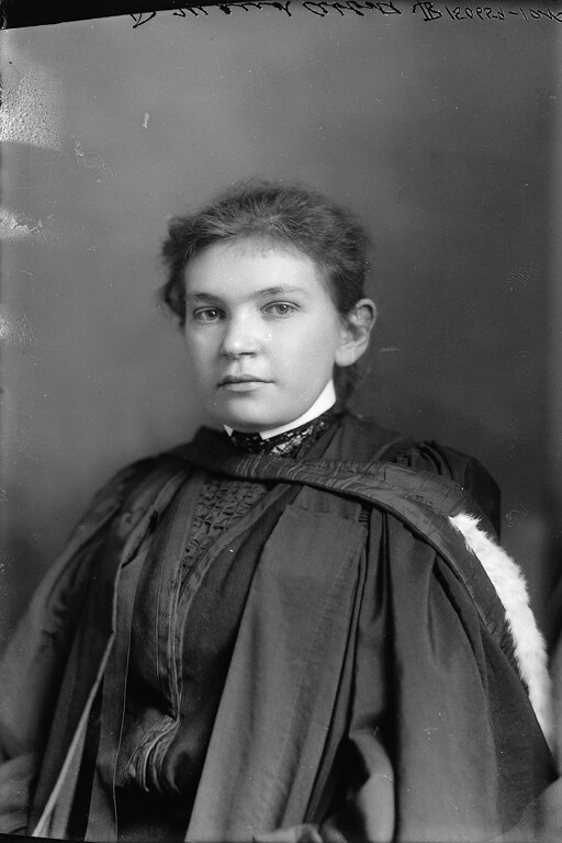
Abbott was born in Saint-André-d'Argenteuil, Quebec, in 1868. She graduated from McGill in arts in 1890, but the university's medical faculty would not admit women. She instead studied at Bishop's College, earning her medical degree in 1894 along with prizes in anatomy and final examinations. Postgraduate study in European clinics and laboratories followed.[2][5]
It is tempting to let rejection determine the rest of this story: a woman barred from one medical school finds a side door into medicine. That framing is true but incomplete. Abbott actively made the museum into an intellectual center. Appointed assistant curator at McGill in 1898, she studied how American teaching museums were organized, took full charge of the collection in 1901, and rebuilt a disordered room into a working relationship among objects, clinicians, and students.[1][2]
One early object showed why organization mattered. Abbott found an unusual heart carrying a label for ulcerative endocarditis but almost no supporting history. William Osler identified it as a specimen donated in 1823 by McGill's first medical dean, Andrew Holmes; it had a single left ventricle. Abbott researched the case and published it in 1901. The jar had survived for nearly eight decades, but preservation alone had not secured its meaning. A specimen became useful only when anatomy, provenance, and clinical interpretation could be reconnected.[1]
Her proposed catalogue grouped material by organ system and paired specimen descriptions with case histories and medical discussion. The same logic shaped her teaching. Informal demonstrations began in 1901; by 1904, the graduating class had thanked her and museum demonstration had entered the curriculum. Students came in groups of about 20, examined specimens and related slides, wrote descriptions, and answered questions in a method Abbott called “descriptive and quizzing.” The point was not to admire a rarity in glass. It was to learn how visible form could discipline a diagnosis.[1][2]
Four hundred cases became more than one thousand
In 1905 Abbott sent Osler a draft catalogue section on diseases of the inner lining of the heart. He invited her to write the congenital-heart chapter for the 1908 edition of his medical reference work. She reviewed the clinical and pathological literature for roughly 400 cases, treating the subject statistically as well as anatomically. She kept adding cases; the chart in her 1936 atlas would contain more than 1,000.[1]
Scale changed the question. A single postmortem heart could show an opening between chambers, an obstructed outflow, or great vessels in an unexpected arrangement. A collection could show which features traveled together, which outward symptoms corresponded to which internal structures, and where apparently similar cases separated. Abbott's atlas made morphology into a comparative language.
The work continued through an institutional reversal. When McGill reorganized pathology and moved its pathological museum in 1924, Abbott lost day-to-day control of accessioning, specimen preparation, and much of the teaching she had built. She spent two years at the Woman's Medical College of Pennsylvania, then returned to Montreal as a research professor and curator of a reduced “Central” Medical Museum. She nevertheless continued gathering congenital-heart specimens from McGill hospitals and colleagues elsewhere.[1][2]
The 1932 London exhibition was therefore more than a late-career display. It compressed a dispersed research practice into a room-sized sequence. After the meeting, Montreal physician David Seecof suggested turning the panels into a book. Abbott dismantled and reorganized them, added cases and explanatory material, and arranged the result into sections on development, comparative anatomy, and clinical disease. The American Heart Association published the atlas in 1936, the year she retired.[1][3]
A map before the route to treatment
Calling the atlas foundational can blur chronology. Abbott did not devise open-heart surgery, and her plates could not provide the moving, living views later offered by cardiac catheterization, echocardiography, or cross-sectional imaging. Much of her evidence came from autopsy. The atlas primarily answered a prior question: what forms of congenital cardiac disease exist, how can they be distinguished, and what clinical histories belong beside them?
That prior question was essential. Operative treatment requires more than the knowledge that a child is cyanotic or that a murmur is present; it requires a sufficiently precise account of the structure that blood is moving through. Abbott's synthesis helped establish the morphology and proposed developmental relationships of congenital heart defects before the later twentieth century made many of them surgical problems.[1]
The surviving collection also shows that an atlas is never the last word. Eighty specimens from Abbott's cardiac collection remained at McGill when they were digitally photographed in 2003–2004. A current museum display presents 39 of them, placing the old catalogue language beside terminology adopted by today's international congenital-heart nomenclature and the International Classification of Diseases. The physical heart may stay fixed in preservative; the categories around it continue to be tested and revised.[1][4]
The unnamed patients inside the classification
There is another boundary to keep in view. McGill's museum states that its older organs—many obtained at autopsy from the 1880s through the 1920s—were generally preserved without specific consent from patients or families. At the time, teaching hospitals treated admission as a kind of assumed permission, a practice medicine has abandoned.[6]
That does not erase the knowledge Abbott assembled, but it changes how the collection should be narrated. Her atlas was made possible by people whose names often receded while their hearts became exemplary cases. The modern museum's memorial language asks viewers to acknowledge those people, not only the curator who made their anatomy legible. A responsible history can hold both facts together: Abbott built an unusually powerful comparative archive, and the archive inherited the consent failures of its medical world.[6]
Abbott died in 1940 before completing a planned larger work on treatment and prognosis. By then, however, she had demonstrated something durable about clinical knowledge. Evidence does not become a field merely by accumulating. It needs an arrangement that lets one case speak to another, a vocabulary that can be corrected, and a teaching practice that makes observation reproducible.
The 32-foot wall in London was the clearest physical expression of that method. Readers did not encounter one spectacular heart. They walked along differences, recurrences, and relationships. Before surgeons could routinely change the anatomy of congenital heart disease, Maude Abbott changed the conditions under which doctors could see it.[1][3]
Sources
- Richard Fraser, “Maude Abbott and the Atlas of Congenital Cardiac Disease,” Cardiovascular Pathology 15 (2006) — professional chronology, case counts, teaching method, exhibition development, and the atlas's medical significance.
- Maude Abbott Medical Museum, McGill University, “Maude Abbott” — education, curatorship, teaching, institutional changes, and career timeline.
- Maude Abbott Medical Museum, McGill University, “Atlas” — dimensions and contents of the 1932 London display and its conversion into the 1936 atlas.
- Maude Abbott Medical Museum, McGill University, “Cardiovascular Collection” — surviving specimens and their presentation alongside current congenital-heart nomenclature.
- Margaret Gillett, “Abbott, Maude Elizabeth Seymour,” Dictionary of Canadian Biography, vol. 16 — biographical record, education, and early barriers in medicine.
- Maude Abbott Medical Museum, McGill University, “In Memoriam” — the provenance of historical teaching specimens and the absence of modern specific consent.
- Wikimedia Commons, “Maude Abbott.jpg” — 1904 Wm. Notman & Son portrait from the McCord Stewart Museum, accession II-150659.