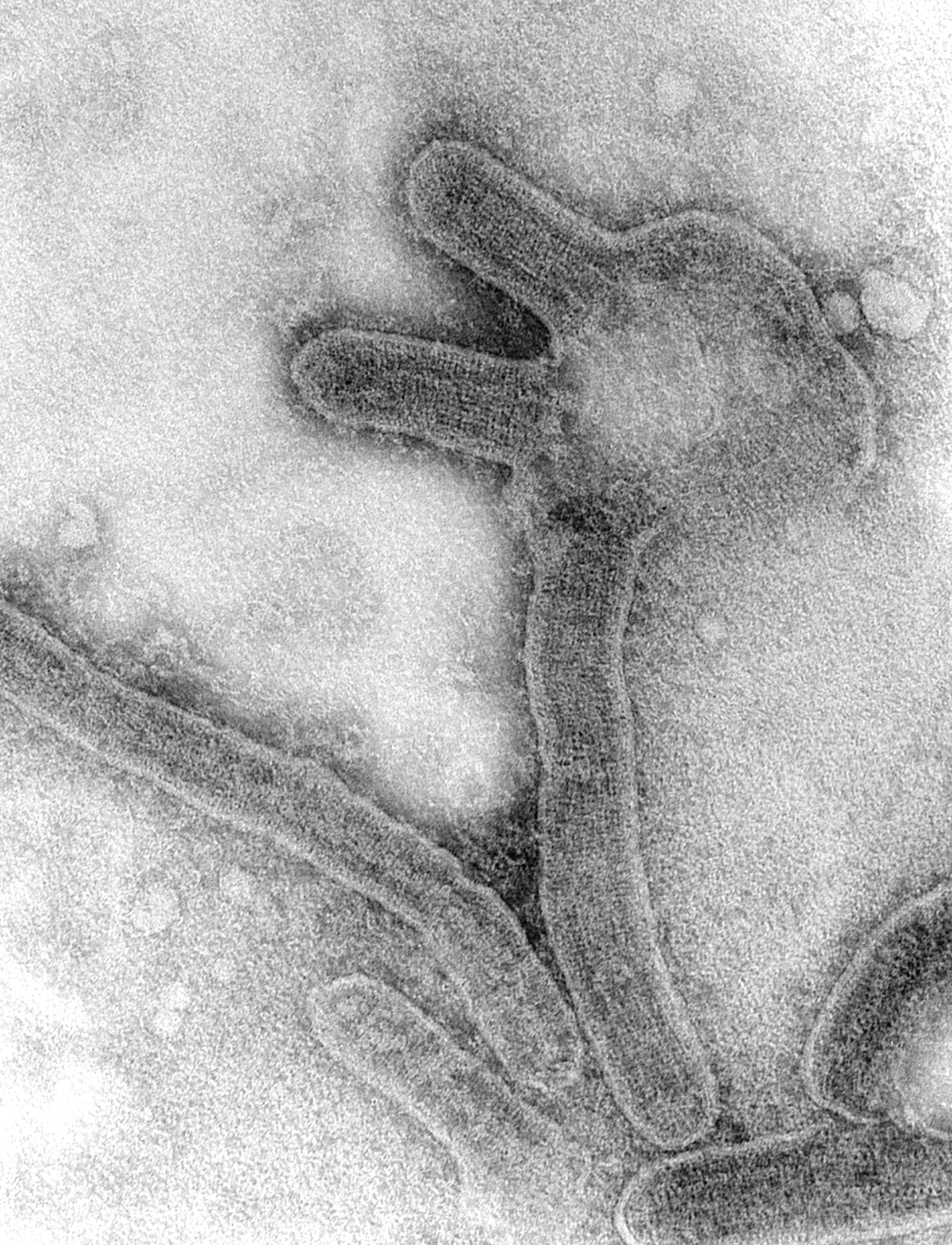
The virus in the cover photograph bends like loose rope. F. A. Murphy made the negative-stain electron micrograph in 1968, after an unfamiliar illness had moved through laboratories and hospitals in three European cities. The image is evidence of what scientists eventually learned to see, not what first alerted them. Before anyone had a recognizable particle, investigators had a pattern of jobs, shipments and touch.[4][8]
In August and September 1967, workers became ill in Marburg an der Lahn and Frankfurt am Main in West Germany, and in Belgrade, Yugoslavia. The current CDC chronology records 31 reported cases and seven deaths, plus one additional primary infection diagnosed retrospectively by serology. A widely used scientific review divides the 31 into 25 primary and six secondary infections. Older reports give slightly different totals because they froze the ledger at different moments.[2][5][6]
The sharp question is not simply how a new virus was discovered. It is how three clusters became one outbreak before the agent had a settled name. The answer began upstream of the microscope: with African green monkeys, the tissues removed from them, and the records that connected laboratories separated by hundreds of kilometres.
The first useful map was a workbench
The three sites did not initially look like one place. Behringwerke AG in Marburg was producing poliomyelitis vaccine. The Paul Ehrlich Institute in Frankfurt was testing vaccine safety. The Torlak Institute in Belgrade also worked on viruses, vaccines and sera. What joined them was a trade in imported grivets—African green monkeys—from the same Ugandan exporter. Workers in the primary cases had handled the animals or their blood, organs and cell cultures; several later infections followed care of sick people or contact with infectious material.[1][2]
The kidneys mattered because primary monkey cells were then used to grow poliovirus vaccine strains. That fact is easy to distort. The documented human outbreak was an occupational and clinical exposure event during animal necropsy, tissue processing and patient care. The cited investigations do not show finished polio vaccine transmitting Marburg virus to vaccine recipients. That is an inference boundary worth stating plainly: an upstream hazard in a manufacturing system is not automatically a contaminated final product.[1][2][3]
Once investigators compared workplaces rather than addresses, coincidence became difficult to sustain. Employees in three cities had not shared a canteen or neighbourhood. Their facilities had shared an animal source and a kind of intimate tissue work. The clinical resemblance supplied one line of evidence; the common exporter and task supplied another. Neither line alone identified a virus, but together they reduced a continental mystery to a traceable exposure network.[1][2]
Belgrade preserved the chain almost hand to hand
The later archive-based reconstruction of the Belgrade episode preserves the supply chain at unusually fine resolution. Between July 18 and August 1, 1967, Torlak received three shipments, each intended to contain 100 grivets. The first and third arrived through London; the second came through Munich. Twelve animals died in transit. Among the 288 that arrived, deaths during quarantine reached 46 of 99, 20 of 95 and 30 of 94 in the three shipment groups.[1]
Those animal deaths were a warning, but not a molecular diagnosis. On August 25, a 45-year-old Torlak veterinarian examined two dead grivets from the third shipment. Internal records reviewed decades later say blood contaminated the outside of a dish. After removing his protective equipment, washing and showering, he handled the dish without gloves and noticed blood on his palm. An earlier published account instead proposed entry through a small abrasion on an unprotected forearm or through the eye. Both accounts locate the hazard in contact with tissue or blood; they disagree about the final centimetres of transmission.[1]
On September 1, the veterinarian developed fever and chills. Conjunctivitis, severe headache, diarrhoea, a spreading rash, low white-cell and platelet counts, liver injury and bleeding followed. He entered the University Clinic of Infectious Diseases on September 7. His illness was not simply “haemorrhage”: dehydration, electrolyte loss and injury across several organ systems were central to the clinical crisis. He recovered after prolonged supportive care, but the case supplied a detailed timeline from a recorded workplace incident to a severe new syndrome.[1]
His wife, who cared for him at home, became ill on September 11. Records describe her contact with soiled linen and blood-stained gauze on September 4 or 5, but accounts differ over whether she touched the material directly. She too recovered. The second case showed that the outbreak map did not end at the animal room: it extended into household care. The surviving record cannot identify the single contact that infected her.[1]
This uncertainty is not a flaw to polish away. The strongest conclusion is that contaminated blood or another close-care exposure infected her during her husband's early illness; the record does not justify choosing among the reported care encounters. More complete contemporaneous documentation of who touched what, or preserved diagnostic samples tied to those encounters, could have shifted the assessment. The surviving evidence described in the cited reconstruction does not resolve it. The honest conclusion is narrower: the secondary case fits direct-contact transmission, while the exact contact remains unproved.[1]
The cases aligned before the agent did
Across the three cities, primary infections clustered among people doing the closest tissue work. Secondary infections were far fewer and occurred around patient care, pathological examination or household contact. That distribution argued against a freely spreading respiratory epidemic in the surrounding cities. It also showed why ordinary hospital work could extend an outbreak whose first boundary appeared industrial.[1][2][5]
The numbers evolved with the investigation. A U.S. National Communicable Disease Center report in June 1968 described 30 cases and seven deaths; the current CDC table lists 31 reported cases, an additional retrospectively detected primary infection and the same seven deaths. The difference is not license to add every historical figure together. It shows how case definitions and late serology can move the edge of an outbreak after its visible transmission has ended.[5][6]
Belgrade's institutional response also began before full identification. Soon after the veterinarian entered hospital on September 7, Torlak halted ongoing vaccine production, removed sick or suspect animals from the surviving groups and extended observation of the remainder. West German and Yugoslav specialists exchanged clinical information and material; an international emergency panel first met in London on October 10. These actions followed the exposure hypothesis—the shared animals and tissue work—not a completed taxonomy.[1]
That sequence is the practical core of the reconstruction. Public health did not need to wait for a perfect name before narrowing contact, stopping the implicated work and coordinating across borders. It did need to preserve enough specimens and records for the etiologic claim to catch up.
A wrong answer arrived before the filament
Early uncertainty produced competing laboratory explanations. One report attributed the illness to a rickettsial or chlamydial agent; later reviewers note that this incorrect claim has sometimes been mistaken for the first identification. Meanwhile, teams in Marburg and Hamburg isolated and characterized a new virus in less than three months. Their 1967 paper described the isolation, identification and structure of an agent from a previously unknown human infection associated with monkeys. A U.S. team subsequently confirmed that the agent recovered from patients was viral and described cylindrical particles 90–100 nanometres wide and 130–2,600 nanometres long.[2][3][4]
Electron microscopy made the strangeness visible. The long particles did not resemble the more compact viral forms many investigators expected; the family name Filoviridae would later draw on filum, Latin for thread. But shape was not a verdict by itself. The persuasive case joined morphology to isolation from patients, antibody responses and the exposure network already traced through the monkey shipments. Epidemiology said where to look; laboratory work said what had been found.[1][2][4]
The cover micrograph must be read with the same restraint. Murphy photographed Marburg virus grown in tissue culture in 1968. It is a real archival laboratory image, but it does not portray the original dish in Belgrade, prove which animal infected which worker or resolve where the grivets acquired the virus. Its value is more exact: it records the physical form of the agent that the outbreak investigation had separated from its early impostors.[4][8]
The shared shipment was proven more strongly than its origin
Investigators could trace the affected European facilities to the same Ugandan exporter. They could also show unusually high illness and death among imported animals and detect antibodies in survivors. Yet shipping routes varied, animals paused in London or Munich, and investigators could not establish that the two grivets examined on August 25 were infected; virus-isolation attempts in tested animals were unsuccessful. Later ecological work makes infection in Uganda plausible, but the 1967 chain cannot establish precisely where or how the grivets acquired the virus.[1][2]
Two interpretations therefore remain at the source end. The leading one is that already infected animals left Uganda and carried the virus into European laboratories. A narrower alternative allows infection or amplification during transport and holding, where animals of different origins could be placed near one another. Preserved pre-shipment specimens or genomic sequences from each link could distinguish them. Belgrade's locally held samples were later destroyed, no Belgrade genome was determined, and no documented specimen chain spanning every link is known to survive. The evidence strongly identifies the shipment system as the vehicle; it does not reconstruct every infection inside it.[1]
The episode became a reference point in the evolution of nonhuman-primate quarantine. A later review of U.S. practice explicitly treats Marburg as evidence that recently imported animals could pose a human health risk and links that recognition to national rules adopted in 1975: registered importers, shipment records, isolation and reporting of suspected illness in animals or workers.[7] It would be too neat to say one outbreak wrote the whole regime. Tuberculosis, yellow fever and other zoonoses were already shaping quarantine. Marburg made the cost of gaps unmistakable.
The city gave the virus its name because Marburg recorded the most cases. The event itself had a different geography. It ran from capture and export, through transit and quarantine, into necropsy rooms, tissue cultures, wards and a household; then it moved again through specimens, phone calls and scientific papers. The filament in the microscope was new. The method that found it was durable: map the work, connect the shipments, act on the exposure and keep the uncertainty visible.
Sources
- Elizabeta S. Ristanović et al., “A Forgotten Episode of Marburg Virus Disease: Belgrade, Yugoslavia, 1967,” Microbiology and Molecular Biology Reviews 84, no. 2 (2020) — archive-based reconstruction of the shipments, two Belgrade cases, containment, source dispute and cross-border response.
- Kristina Brauburger et al., “Forty-Five Years of Marburg Virus Research,” Viruses 4, no. 10 (2012) — synthesis of the 1967 epidemiology, case classification, identification timeline and later ecological boundary.
- Rudolf Siegert, Hsin-Lu Shu, Werner Slenczka, Dietrich Peters and Günther Müller, “Zur Ätiologie einer unbekannten, von Affen ausgegangenen menschlichen Infektionskrankheit” [On the cause of a previously unknown human infection transmitted from monkeys], Deutsche Medizinische Wochenschrift 92 (1967) — the original report of isolation, identification and structure of the new agent.
- Robert E. Kissling, R. Q. Robinson, Frederick A. Murphy and S. G. Whitfield, “Agent of Disease Contracted from Green Monkeys,” Science 160 (1968) — confirmation of viral character and electron-microscopic measurements.
- U.S. National Communicable Disease Center, “Follow-Up Obscure Disease Related to African Monkeys,” Morbidity and Mortality Weekly Report, June 15, 1968 — contemporaneous case count, clinical summary and laboratory characterization.
- Centers for Disease Control and Prevention, “History of Marburg Outbreaks” (updated June 23, 2026) — current outbreak ledger and treatment of the retrospectively diagnosed 1967 case.
- Jeffrey A. Roberts and Kirk Andrews, “Nonhuman Primate Quarantine: Its Evolution and Practice,” ILAR Journal 49, no. 2 (2008) — Marburg's place in the development of U.S. importation, quarantine and reporting rules.
- CDC Public Health Image Library, image 7218 — archive record for F. A. Murphy's black-and-white 1968 transmission electron micrograph used as the cover image.