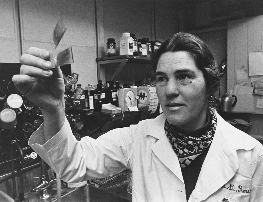
At Janet Rowley's dining table, photographs became loose pieces of evidence. She cut individual chromosomes from laboratory prints and arranged them in pairs. Her children joked about the paper dolls. The University of Chicago's account places this work in her Hyde Park home in 1972, after she had returned from Oxford with new methods for making chromosomes distinguishable.[1]
The table is a memorable setting, but the question occupying it matters more. Leukemia cells could contain conspicuously abnormal chromosomes. Were those abnormalities merely damage left by the disease, or could a particular alteration help explain it? Rowley's achievement was to make the question more precise: look for the same change in different patients, then ask what that change actually does.
Learning to recognize the pieces
Rowley's route to research included delays and part-time work. In the National Library of Medicine's biographical interview, she recalled that a quota for women postponed her entry to medical school by nine months. She earned her medical degree in 1948, worked in public-health clinics and subsequently at a Chicago clinic for children with developmental disabilities, and raised four sons while continuing her career.[2]
Chromosome research gave that clinical background a new direction. Following training in England, she returned to Chicago in 1962 to work in hematology. A later Oxford visit, in 1970–71, introduced the banding methods she would use on leukemia samples.[1][2] Her skill depended on learning how to see differences that an earlier preparation could conceal.
The problem was identification. Before banding, several chromosomes looked sufficiently alike to be classified together by size and shape. An extra member of a group did not tell a researcher which chromosome had multiplied. Distinctive bands made a more exact comparison possible. In their 2011 symposium account, Rowley and fellow investigators described how this allowed her to revisit earlier observations and recognize recurring changes.[3]
That altered the value of repetition. Finding an extra chromosome in two patients might once have meant two vaguely similar abnormalities. Identifying the same chromosome in both made the observation more specific. A better stain did more than sharpen a photograph; it changed what could count as matching evidence.
The chromosome that looked too short
Peter Nowell and David Hungerford had reported the Philadelphia chromosome in 1960, establishing a recurring chromosome abnormality in chronic myelogenous leukemia, usually shortened to CML.[3] Rowley therefore entered an investigation already under way. Her contribution would change the explanation of a familiar object.
Her 1973 Nature letter described cells from nine consecutive patients. Chromosome 22 appeared shortened. But the new staining methods also exposed additional material at the end of one chromosome 9. Its amount approximately matched what appeared absent from 22. The finding suggested that material had moved between chromosomes.[4]
The important move was to read the two observations together. A short chromosome invited a story about loss. A short chromosome accompanied by an extended partner invited a story about relocation. Instead of treating every malformed chromosome as an isolated specimen, Rowley compared the whole set.
The paper's language was appropriately provisional. Its abstract proposed a previously undetected translocation—a movement of chromosome material—rather than claiming to have identified the responsible genes or explained their activity.[4] The visible exchange supplied a location for further investigation. It could not, by itself, reveal the molecular machinery operating there.
Comparing stages of the disease
Rowley's later recollection adds an essential comparison. She had been examining cells from CML's advanced blast phase, where numerous abnormalities could complicate interpretation. She returned to preparations from the chronic phase and found the suspected exchange there too. She also examined stimulated T cells from several patients and found normal chromosomes in those cells.[3]
Each comparison narrowed the explanation. An alteration already visible in chronic-phase samples could not be dismissed simply as wreckage appearing only during the terminal transformation. Normal findings in another cell population challenged the idea that this was just a chromosome variant present throughout the patient's body. Neither observation alone proved how leukemia began. Together, they made the proposed rearrangement harder to explain away.
This is the less picturesque work behind the dining-table story: selecting comparisons that could expose a mistaken interpretation. Persistence mattered because it brought additional evidence to the argument. Repeating a conviction would have accomplished much less.
From an address to a mechanism
Later teams investigated the genes at the exchange. In 1985, Emma Shtivelman and colleagues reported a fused RNA transcript joining BCR sequences to ABL sequences. They proposed that the resulting fused protein contributed to malignancy.[5] The chromosome observation now had a molecular counterpart: a new combination of genetic instructions.
A different kind of test followed. In 1990, George Daley, Richard Van Etten and David Baltimore introduced a gene encoding P210 BCR–ABL into mouse bone marrow cells, then transplanted those cells into recipient mice. The animals developed several blood malignancies, including a syndrome resembling human chronic-phase CML.[6] This was experimental evidence that the fusion could drive disease, with the limits of a mouse model and more than one resulting malignancy.
The distinction between these studies is central to Rowley's legacy. Chromosome microscopy identified a recurrent rearrangement. Molecular analysis characterized its product. Experimental manipulation tested what that product could cause. Those achievements supported one another, while answering different questions. Assigning the entire chain to a single discovery would erase the work that made the original observation useful.
Treatment required another test
By 2001, Brian Druker and colleagues could report a phase 1 study of STI571, the drug known as imatinib, which inhibits BCR–ABL's kinase activity. Their study enrolled 83 patients with chronic-phase CML after interferon-alpha treatment had failed. Among 54 patients receiving at least 300 milligrams daily, 53 achieved a complete hematologic response; seven achieved complete cytogenetic remission.[7]
Those outcomes require careful reading. A hematologic response concerns the blood picture; a cytogenetic response concerns the chromosome abnormality in examined cells. The striking early results did not mean that 53 people had been cured. This was an early dose-escalation study in a defined patient group, rather than a randomized demonstration of long-term survival benefit.[7]
It did show why identifying a disease's molecular dependency could matter to treatment. The drug tested an intervention against the abnormal protein's activity. Rowley's rearrangement had helped direct investigators toward that protein, but developing and testing the inhibitor required a further body of work.
Her surviving papers preserve a similarly broad view of research. The University of Chicago Library's finding aid lists correspondence, laboratory records, drafts, notes, photographs and publication files alongside awards.[8] A celebrated result sits within an accumulation of working material.
The enduring image is therefore worth keeping in focus: a physician arranging cut photographs so that one patient's chromosomes could be compared with another's. The small pieces mattered because their relationships could be checked. Rowley helped turn an abnormal shape into a question that other scientists could pursue, refine and eventually act upon.
Sources
- University of Chicago News, “Janet Rowley, cancer genetics pioneer, 1925–2013” (December 17, 2013)—Oxford training, laboratory work and the dining-table account.
- National Library of Medicine, “Biography: Dr. Janet Davison Rowley,” Changing the Face of Medicine—career history and Rowley's recollections of medical training.
- H. Sharat Chandra and colleagues, including Peter Nowell and Janet Rowley, “Philadelphia Chromosome Symposium: commemoration of the 50th anniversary of the discovery of the Ph chromosome,” Cancer Genetics 204 (2011), 171–179—participant recollections of identification methods and comparison samples.
- Janet D. Rowley, “A New Consistent Chromosomal Abnormality in Chronic Myelogenous Leukaemia identified by Quinacrine Fluorescence and Giemsa Staining,” Nature 243 (1973), 290–293—original report; the linked abstract describes the nine-patient series and proposed translocation.
- Emma Shtivelman and colleagues, “Fused transcript of abl and bcr genes in chronic myelogenous leukaemia,” Nature 315 (1985), 550–554—original molecular study and abstract.
- George Q. Daley, Richard A. Van Etten and David Baltimore, “Induction of chronic myelogenous leukemia in mice by the P210bcr/abl gene of the Philadelphia chromosome,” Science 247 (1990), 824–830—experimental mouse study, PubMed abstract.
- Brian J. Druker and colleagues, “Efficacy and safety of a specific inhibitor of the BCR-ABL tyrosine kinase in chronic myeloid leukemia,” New England Journal of Medicine 344 (2001), 1031–1037—phase 1 study, eligibility and response outcomes, PubMed abstract.
- University of Chicago Library, Guide to the Janet D. Rowley Papers, 1940–2013—collection scope and organization; cited as a finding aid, not as an inspection of the underlying files.
- University of Chicago Library, “Rowley, Janet,” Archival Photographic Files, apf7-01134—Chicago Maroon photograph of Rowley in her laboratory, dated to the 1980s.