
A dinosaur skeleton contains no reading for kilograms. Bone can preserve the dimensions of a load-bearing column, but not the flesh that once surrounded it. A mounted skeleton can preserve a whole-body geometry, but its empty spaces do not say how much muscle filled the thigh, how wide the abdomen was, or how much air reduced the density of the neck and trunk. Yet body mass is the quantity researchers need before they can ask many larger questions about locomotion, growth, metabolism, reproduction, and ecology.[3][4]
Paleontologists have therefore built two kinds of scale. One starts with living animals of known mass and asks how the circumferences of their major limb bones change as bodies become heavier. The other rebuilds an extinct body around a skeleton, calculates its volume, and assigns densities to its tissues and respiratory spaces. The methods are often described as competitors. A large review of dinosaur estimates found them more useful as checks on one another: one carries empirical calibration from living animals, while the other retains the extinct animal's particular shape.[3]
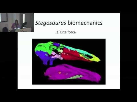
The two videos in this collection show different parts of that argument. A University of New England presentation lays out the two mass-estimation traditions and the assumptions behind each.[1] A Natural History Museum Science Forum seminar by Charlotte Brassey then puts both methods to work on the same unusually complete Stegosaurus skeleton.[2][8] Watched together, they reveal that “weighing” a dinosaur is not one calculation. It is a chain from specimen to measurement, from measurement to model, and from model to a range that should remain open to revision.
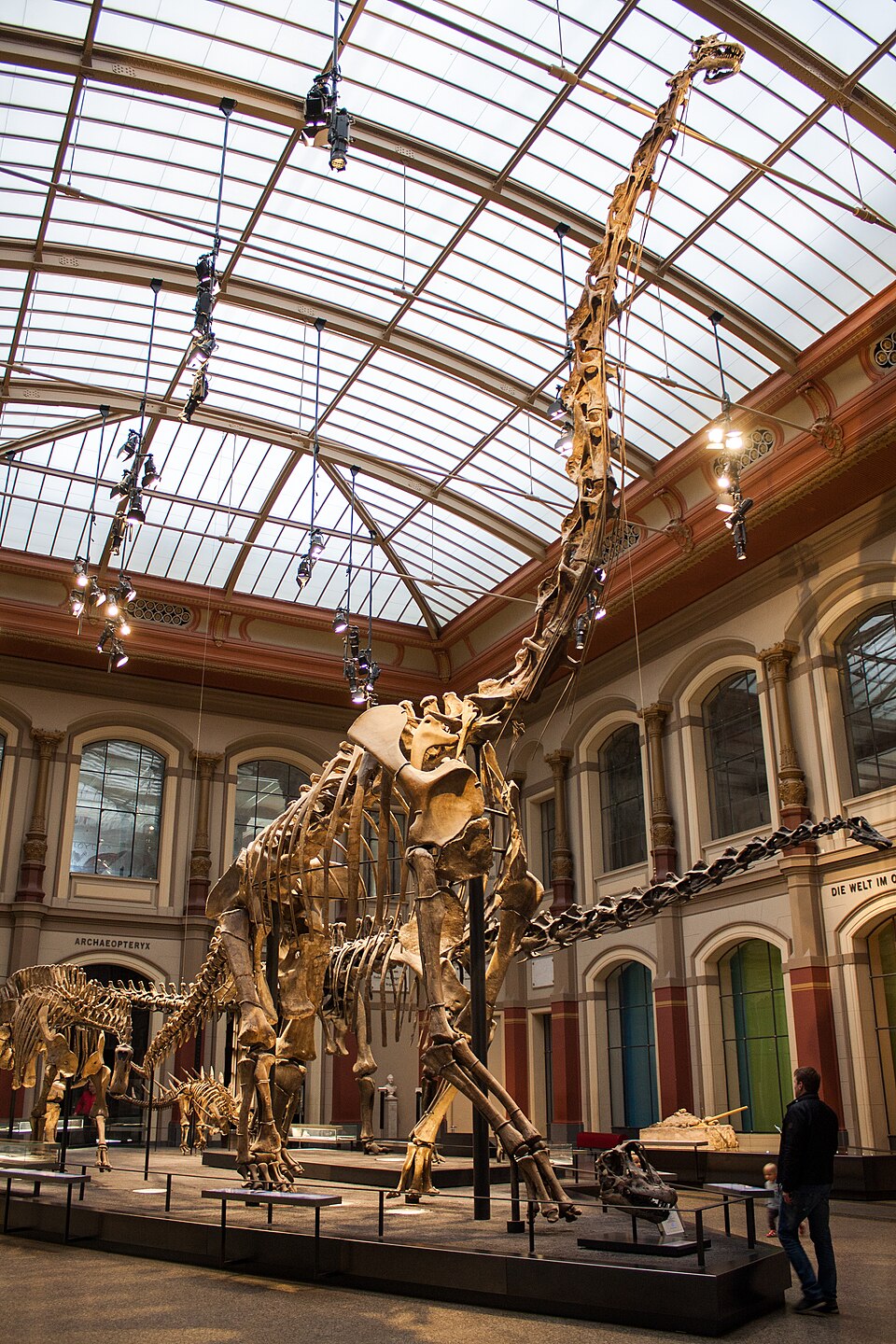
Image context: the cover photograph shows the Berlin mount of Giraffatitan brancai, the same reconstructed skeleton scanned for a 2012 minimum-convex-hull study. The animal towers over the gallery, but the relevant scientific object is more exacting than its silhouette: the articulation of every mounted bone determines the digital envelope built around it.[6][9]
1. The bone scale measures a history of carrying weight
Nicolás Campione's 28-minute UNE Discovery presentation begins with the biological importance of size, then separates the field into its two main approaches around 5:45: limb-bone scaling and reconstruction.[1][3] The chapters on minimum shaft circumference and reconstructed volume follow at about 7:08 and 7:25.[1] That division is the video's most useful feature. It keeps a familiar museum question—“How heavy was it?”—from collapsing into a single clever trick.
The limb method begins with a mechanical fact that living animals have already tested: the major bones nearest the body must repeatedly transmit its weight. In terrestrial quadrupeds, the combined minimum shaft circumferences of the humerus and femur have a highly conserved relationship with mass. Campione and Evans assembled measurements from living mammals and reptiles and found that this relationship held despite large differences in gait, limb posture, and evolutionary history.[4] The equation is not reading fossilized weight directly. It is applying a relationship calibrated on bodies whose weights and bones can both be measured.
Bipedal dinosaurs require an additional step because their forelimbs do not share the standing load. A later study converted the quadrupedal relationship to the geometry of one principal weight-bearing pair, allowing femoral circumference to be used within the same empirical framework.[5] This matters because “use a big leg bone” would be a poor method. The defensible version specifies which circumference is measured, how the living comparison was fitted, whether the animal was bipedal or quadrupedal, and how uncertainty in the regression follows the estimate.
The attraction is breadth. A reasonably preserved humerus and femur can support an estimate even when there is no complete torso to reconstruct. That makes extant scaling practical for comparing many taxa across a lineage or an ecosystem.[3] Its weakness is the same economy. One or two bones may be unusually robust or gracile for reasons that a general equation cannot fully capture. A fossil may also sit beyond the body sizes represented by the living calibration sample. The result can be empirically grounded yet imprecise for a particular individual.
The presentation's reconstruction section supplies the complementary question: what if the rest of the skeleton contains information that a limb circumference throws away?[1] A long neck, deep trunk, heavy tail, or unusually light pneumatic skeleton can redistribute volume and mass without changing one measured shaft in a predictable way. Whole-body models preserve those differences. They also introduce decisions about missing anatomy, posture, flesh, and density. Neither pan of the scale is empty of assumptions; the assumptions simply enter at different places.
2. Sophie puts both estimates on the same skeleton
Brassey's Natural History Museum seminar supplies the comparison that a general methods lecture cannot: two answers for one individual.[2] The subject is NHMUK R36730, the Stegosaurus stenops nicknamed Sophie, from the Upper Jurassic Morrison Formation near Shell, Wyoming. All body regions are represented except the left forelimb and part of the tail, and histology identifies the animal as a young adult that was still growing.[8] The talk moves from photogrammetric digitization near 1:22 to body mass around 17:25, then makes the modeling choices visible through “digital plasticine,” convex hulls, and sensitivity analysis from roughly 19:33 onward.[2]
The team's photogrammetric models preserved the form of Sophie's disarticulated bones, but they did not arrive as a finished animal. The researchers posed the skeleton digitally, then changed intervertebral spacing, rib flare, and shoulder-blade position to produce minimum, preferred, and maximum-volume articulations. Convex hulls wrapped functional units such as the trunk, neck, tail, and limbs; dermal armor was calculated separately.[8] This is the boundary the video makes tangible. Digitization can capture fossil geometry. It cannot decide articulation, missing anatomy, or the distribution of living tissue without anatomical judgment.
The preferred convex-hull model yielded 1,560 kilograms, with a 95% prediction interval of 1,082–2,256 kilograms. Changing the articulation moved the central estimates from 1,311 to 1,894 kilograms.[8] Limb-circumference equations initially gave substantially higher values, from 2,355 to 3,751 kilograms. For the preferred body to reach the upper limb-based estimate, its modeled volume had to expand by 165%, producing a visibly excessive soft-tissue envelope.[8] At that stage, the methods looked incompatible.
Growth stage changed the diagnosis. The standard limb equations had effectively treated Sophie's robust limbs as though they belonged to a fully adult reference. When the authors used known adult Stegosaurus specimens as endpoints and scaled back to Sophie's femur length, the limb-based estimates fell to 1,823 and 2,158 kilograms—inside the volumetric intervals.[8] The discrepancy was useful because it pointed toward ontogeny. A “winning” method would have hidden the most interesting result: the skeleton was complete enough to show that age, not merely fleshiness, separated the first answers.
Digital models can also ask questions that a total mass alone cannot. In one influential workflow, researchers laser-scanned mounted dinosaur skeletons, divided the virtual animals into segments, drew body envelopes around those segments, assigned bulk densities, subtracted respiratory spaces, and calculated center of mass and rotational inertia.[7] Because the models were digital, the team could make slimmer and fuller versions and vary the sizes of air sacs. The range was not a failure to pick the “right” silhouette. It exposed how much the result depended on anatomy that fossil bone does not preserve.
The Berlin Giraffatitan in the cover photograph became the subject of a more automated volumetric experiment. Sellers and colleagues wrapped laser-scanned skeletons in minimum convex hulls—the tightest convex envelopes able to contain their points. Tests on 14 mounted mammals showed that these bare-bone hulls consistently fell below living body mass, so the researchers derived an empirical expansion from the mammal sample. Applied to the Berlin mount at an assumed mean density of 800 kilograms per cubic meter, the method produced an estimate of 23,200 ± 2,200 kilograms.[6]
That figure is memorable, but the calibration is the real lesson. The hull alone is deliberately too lean. Its correction came from living mammals, and the authors cautioned that the same expansion might not fit birds, reptiles, long necks, or heavy tails equally well.[6] The mount also has to be accurately reconstructed, especially through the ribs and torso where much of the volume sits. A fast, repeatable algorithm can reduce artistic input after scanning; it cannot remove decisions already embodied in the skeleton's pose or in the choice of living comparison.
Agreement is a test, not permission to erase the range
When Campione and Evans compared the two traditions across published dinosaur estimates, 73% of volumetric reconstructions fell within the 95% prediction intervals of the extant-scaling relationship. Of the volumetric outliers, almost three quarters were below the lower interval.[3] The broad agreement matters because the approaches do not inherit the same weaknesses. Their disagreements matter for exactly the same reason.
A low volumetric estimate may signal a body wrapped too tightly around the skeleton, a density set too low, or a mount with a narrow reconstructed torso. A high limb-scaling estimate may expose an animal whose weight-bearing bones were unusually stout, or a regression stretched beyond its best calibration range. The specimen can complicate both: deformation changes circumferences, missing ribs weaken a body envelope, and a juvenile or exceptionally old individual should not be mistaken for a species average.
This gives every estimate three visible layers. The specimen layer records completeness, association, deformation, and growth stage. The model layer records the living calibration, skeletal articulation, soft-tissue envelope, respiratory volume, and density. The reporting layer distinguishes one reconstructed individual from a typical animal and gives a plausible interval rather than a false weighbridge reading. A headline number without those layers is easier to repeat, but much harder to use.
The videos finally make the two pans of the scale feel less metaphorical. The UNE presentation shows why a circumference can carry information accumulated through millions of steps by a living body.[1] Brassey's seminar shows one skeleton becoming several explicit, testable bodies—and then shows growth stage pulling two apparently incompatible methods together.[2][8] One method asks what load a bone of this size usually bore. The other asks how much modeled matter can plausibly surround this particular skeleton. The strongest estimate is not the one that makes uncertainty disappear. It is the one that lets these independent questions meet—and makes their remaining distance part of the result.
Sources
- UNE Discovery, “How to weigh a dinosaur!”, YouTube video, 2020.
- SciFri Seminars (Natural History Museum Science Forum), Charlotte Brassey, “NHM SciFri Seminar — Project Stegosaur,” YouTube video, January 2015.
- Nicolás E. Campione and David C. Evans, “The accuracy and precision of body mass estimation in non-avian dinosaurs,” Biological Reviews 95 (2020), PubMed record and abstract.
- Nicolás E. Campione and David C. Evans, “A universal scaling relationship between body mass and proximal limb bone dimensions in quadrupedal terrestrial tetrapods,” BMC Biology 10 (2012).
- Nicolás E. Campione et al., “Body mass estimation in non-avian bipeds using a theoretical conversion to quadruped stylopodial proportions,” Methods in Ecology and Evolution 5 (2014), Smithsonian Research Online record.
- William I. Sellers et al., “Minimum convex hull mass estimations of complete mounted skeletons,” Biology Letters 8 (2012).
- Karl T. Bates et al., “Estimating Mass Properties of Dinosaurs Using Laser Imaging and 3D Computer Modelling,” PLOS ONE 4 (2009).
- Charlotte A. Brassey, Susannah C. R. Maidment, and Paul M. Barrett, “Body mass estimates of an exceptionally complete Stegosaurus: comparing volumetric and linear bivariate mass estimation methods,” Biology Letters 11 (2015).
- Shadowgate, “Museum für Naturkunde (36556352434),” 2017 photograph of the Berlin Giraffatitan mount, Wikimedia Commons, CC BY 2.0.