A stylophoran offers few familiar clues: a flattened, plated body and one jointed appendage. For generations of researchers, deciding what that appendage did meant deciding which way the whole animal faced.[1]
The consequential evidence eventually appeared inside it. A Moroccan fossil preserved a branching arrangement of soft tissues, recorded in minerals. Reading those branches gave researchers a way to test the animal's identity against something more discriminating than its outline.[3]
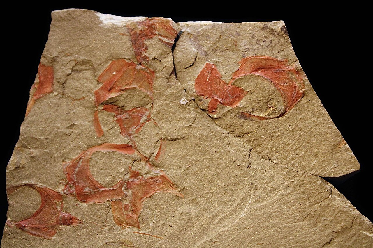
Which end was the front?
Stylophorans lived from the Cambrian into the Carboniferous. Their skeletons carried an important clue: stereom, the microscopic meshwork characteristic of echinoderm plates. Yet they lacked the conspicuous fivefold organization associated with sea stars and sea urchins.[1]
Several interpretations competed. The appendage could be a stalk, a feeding arm, or a tail. Under the influential calcichordate hypothesis, these plated creatures belonged near the ancestry of chordates, the group that includes vertebrates. Treating the appendage as a tail reversed the orientation used by advocates of the feeding-arm interpretation.[1]
That disagreement reached into family trees. In a 2005 analysis, Bertrand Lefebvre showed how assumptions about corresponding skeletal plates could shape the evolutionary relationships recovered by combining earlier trees.[1] Agreement between reconstructions could therefore inherit an anatomical assumption. The productive question was what observation might distinguish those assumptions in the first place.
A branching trace in the stone
The material came from Bou Izargane, about 18 kilometers north of Zagora in Morocco's Anti-Atlas. Here, the Fezouata Formation preserves Early Ordovician marine life, including animals whose softer structures usually disappear. Specimens from the investigation are held at Cadi Ayyad University in Marrakesh.[2]
The fossil-bearing level belongs to the late Tremadocian, an early division of the Ordovician. In the 2019 study by Lefebvre and colleagues, specimen AA.BIZ15.OI.259 preserved two lengthwise canals in its appendage. Small lateral tubes branched from the outer canal and extended between hinged plates.[3]
These were mineral records of tissue. Pyrite had formed in association with the soft parts; subsequent oxidation produced colored iron-rich traces. Elemental mapping helped establish where those traces lay within the skeletal impressions.[2] The anatomy had to be read through the chemistry of its preservation.
The branching canal was interpreted as part of a water vascular system, a fluid-filled network supplying tube feet. The lateral structures were identified as those tube feet. That arrangement fits an echinoderm feeding arm. The external projections and opening plates were incompatible with the enclosed stalk-like anatomy proposed by a competing model.[3]
The inference depends on relationships between parts: where a canal runs, what branches from it, and whether those branches reach outside the body. A long mark alone would be ambiguous. A repeated arrangement that connects internal and external structures gives the comparison much more force.
Why hundreds of fossils could keep the secret
The same specimen appears under different genus names in the literature: Thoralicystis in the 2019 paper, Bohemiaecystis in a 2023 study. Its collection number makes the continuity traceable despite the revised identification.[3][4]
Farid Saleh and colleagues examined 423 stylophorans from a single Fezouata level to investigate their decay. The water vascular system occurred in just one specimen. Other internal remains survived more frequently, particularly the foregut within the proximal, or bodyward, portion of the appendage.[4]
Their explanation turns the skeleton into part of the preservation process. As muscles decayed, the delicate cover plates could open, exposing the tissues beneath. The more resistant enclosure nearer the body protected its contents for longer. Different parts of one carcass therefore offered different chances for mineral replication.[4]
This makes absence difficult to interpret. Counting complete-looking fossils would exaggerate how many opportunities researchers had to examine the crucial anatomy. The relevant unit of evidence is the preserved structure, not simply the number of animals on a slab.
From identity to movement
Recognizing a feeding arm still leaves questions about how an animal used it. In 2020, Elizabeth Clark and colleagues investigated another stylophoran, Phyllocystis crassimarginata, from the Early Ordovician of France. A fossil preserved as a nearly three-dimensional mold allowed them to reconstruct its appendage from micro-CT scans and test the range of motion at its joints.[5]
Their model made one proposed locomotion strategy unlikely: efficient movement driven by up-and-down strokes. Sideways motion of the proximal appendage offered a more plausible possibility for this species.[5]
That result concerns mechanical feasibility. It does not record a living animal's gait, establish how frequently it moved, or supply one behavior for every stylophoran. The model also rests on a different taxon from the Moroccan fossil. Its value here is to keep two questions separate: identifying an appendage's anatomical organization and reconstructing everything it could do.
Look back at the thin extensions on the slab. Their outlines invite an immediate answer—arm, stalk, tail. The Fezouata specimen earned a stronger answer through structures most neighboring fossils had lost. A few branching mineral traces changed the direction in which researchers could read an entire animal.
Sources
- Bertrand Lefebvre, “Stylophoran supertrees revisited,” Acta Palaeontologica Polonica 50 (2005), 477–486—skeletal anatomy, competing interpretations and the influence of anatomical assumptions on evolutionary trees.
- Cadi Ayyad University, “Un débat scientifique finalement clôt grâce au gisement de Fezouata à Zagora, Anti-Atlas Maroc” (February 13, 2019)—locality, mineral preservation, research context and museum collections; in French.
- Lefebvre and colleagues, “Exceptionally preserved soft parts in fossils from the Lower Ordovician of Morocco clarify stylophoran affinities within basal deuterostomes,” Geobios 52 (2019), 27–36—author manuscript, including specimen AA.BIZ15.OI.259 and its canal-and-tube anatomy.
- Saleh and colleagues, “Skeletal elements controlled soft-tissue preservation in echinoderms from the Early Ordovician Fezouata Biota,” Geobios 81 (2023), 51–66—423-specimen study of skeletal disarticulation and selective soft-tissue preservation.
- Clark, Hutchinson, Bishop and Briggs, “Arm waving in stylophoran echinoderms: three-dimensional mobility analysis illuminates cornute locomotion,” Royal Society Open Science 7 (2020), 200191—Phyllocystis joint mobility; full text in the NSF Public Access Repository.
- Prehistorica CM, “Thoralicystis zagoraensis.jpg,” Wikimedia Commons (June 11, 2026)—original photograph of stylophorans from the Fezouata Formation, licensed CC BY 4.0.