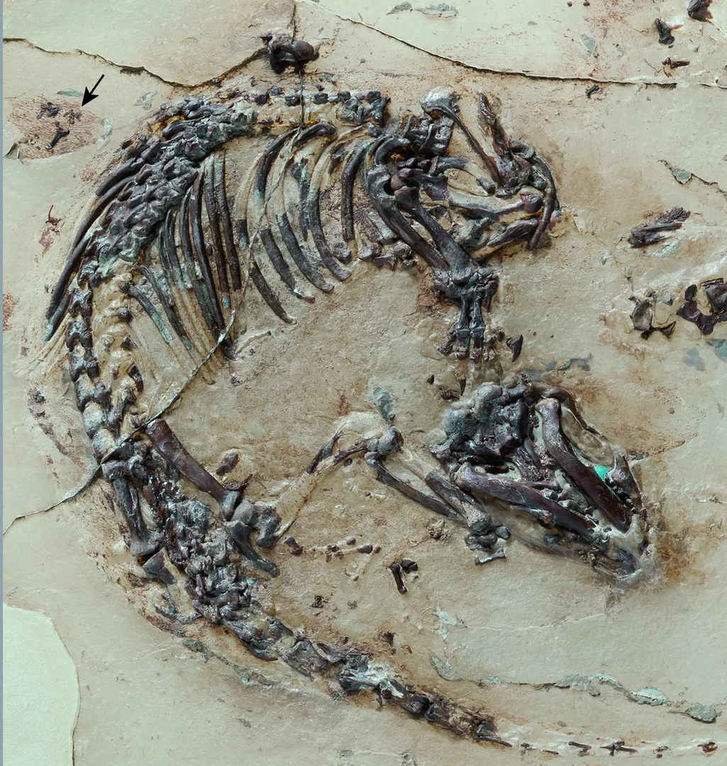
The most revealing part of Spinolestes is too small to appreciate in a whole-fossil photograph. Around its skeleton, the preserved coat resolves under magnification into hairs emerging together from individual pores. A Cretaceous mammal has left evidence of how its fur grew.[1]
Described by Thomas Martin and colleagues in 2015, Spinolestes xenarthrosus comes from Las Hoyas, Spain, and was reported as approximately 125 million years old. It belongs to the extinct eutriconodont mammals. Its covering included ordinary fur, a mane, tiny spines and keratinous plates: several ways of building a skin surface, assembled on one small body.[1]
The temptation is to dress it in familiar names—mouse, hedgehog, miniature armadillo. Reading the specimen closely produces something more interesting. Familiar structures recur in an animal that cannot be assigned to any living mammal group.[2]
Start at the skin
The University of Bonn's photograph shows a prepared object. Researchers removed the fossil from its limestone and transferred it to a synthetic support. That pale background belongs to the specimen's laboratory history; it is not a view of an untouched Cretaceous bedding plane.[2]
This matters when looking at the outline around the bones. A photograph brings the animal within reach, but preparation and magnification determine which features become legible. The fine structure of a coat cannot be read simply by enlarging a mental picture of a furry silhouette.
The University of Chicago's account describes preservation through phosphatic fossilization. Scanning electron microscopy revealed follicles, hair bulbs and details within individual shafts. Multiple hairs could emerge from a single pore, an arrangement called a compound follicle. The researchers also identified unusually shortened hairs consistent with a fungal infection.[3]
The last claim belongs at a different level from observing a pore. A damaged hair is a preserved feature; an infection is a diagnosis made by comparison. It is a plausible glimpse of an individual's condition, without making every irregularity in its coat a medical record.
A spine has a structure before it has a purpose
The original description identifies the small spines as bundles of hair-like tubules. It also records additional articulations between vertebrae in the rear part of the back. Those joints strengthened the spine and resembled arrangements that evolved independently in living xenarthran mammals, the group containing armadillos and anteaters.[1]
This is an animal with several specializations, rather than a generic early mammal waiting for later evolution to give it useful equipment. Yet identifying equipment and reconstructing its use are separate tasks.
Bonn's researchers compared its covering with that of living spiny mice, which can shed spines when seized by a predator. A similar escape function in Spinolestes was a suggestion. They likewise used the reinforced back of an African armored shrew to suggest possibilities for feeding, while acknowledging uncertainty about the fossil mammal's behavior.[2]
These comparisons are productive because they pose testable functional questions. Could a structure resist a particular force? Could it detach? Resemblance offers a starting point; it does not supply an eyewitness account of a Cretaceous encounter.
The organs leave a different kind of outline
Inside the body, researchers recognized microscopic lung bronchioles and iron-rich residues associated with the liver. A curved boundary separated the regions. Their arrangement supported the inference of a muscular diaphragm, the partition involved in mammalian breathing.[3]
That wording deserves care. Recognizing organ traces and using their positions to infer a separating muscle are different evidential steps. A compelling anatomical inference does not require the muscle itself to be displayed as an intact structure.
The fur and the organ boundary therefore reward different kinds of attention. In one, microscopic structures can be examined directly. In the other, the spatial relationship between surviving traces helps reconstruct something less directly accessible. Exceptional preservation improves both inquiries without making them identical.
What a detached hair cannot tell us
A useful comparison comes from a 2020 study of Spanish Cretaceous amber by Sergio Álvarez-Parra and colleagues. Three aligned hair strands from Ariño preserved detailed surface scales, but lacked their tips and follicles. The researchers could identify mammalian hair without confidently identifying the mammal that produced it.[4]
They explicitly compared the strands with Spinolestes. The scale patterns differed, and knowledge of hair in other Cretaceous mammals remained too incomplete to assign the Ariño material securely. Even a resemblance to hairs of living mammals could not bridge that gap.[4]
The contrast explains the value of the Las Hoyas specimen. Detail and connection answer different questions. A finely preserved strand can establish what kind of material it is; a coat associated with skin and a diagnostic skeleton lets researchers place that material on a particular animal.
Looking back at Spinolestes, the indistinct margin around the bones becomes the beginning of an investigation. The photograph brings us to the body. The follicles take us into the construction of its coat. Keeping those observations connected is what gives this small fossil its extraordinary reach.
Sources
- Thomas Martin and colleagues, “A Cretaceous eutriconodont and integument evolution in early mammals,” Nature 526 (2015), 380–384 — original description, compound follicles, skin structures, vertebral anatomy and inferred diaphragm.
- University of Bonn, “Ursäugetier litt vermutlich unter Haar-Erkrankung” (2015; German) — preparation history, Georg Oleschinski's specimen photograph and the researchers' functional comparisons.
- Kevin Jiang, “125 million-year-old fossil reveals early mammalian hair and spines,” University of Chicago News (2015) — microscopy, phosphatic preservation, possible hair disease and organ traces.
- Sergio Álvarez-Parra and colleagues, “Cretaceous amniote integuments recorded through a taphonomic process unique to resins,” Scientific Reports 10 (2020), 19840 — Ariño hair strands, comparison with Spinolestes and limits of taxonomic identification.