Paleopathology has an image problem. The public-facing version often arrives as dinosaur gore: bite marks, smashed frills, infected jaws, broken tails, a fossil biography reduced to one violent moment. The better reading begins later in time. A pathologic bone matters because the animal did not die instantly, because tissue responded, because healing or remodeling had time to leave a trace, and because that trace can be compared to living vertebrate bone with more discipline than the spectacle usually suggests.[1][2][3]
That deep-time perspective is exactly how the field defines itself. The Museum für Naturkunde describes paleopathology as the study of diseases recorded in fossil vertebrate skeletons, but the important part is the question set that follows: what kinds of disease affected extinct animals, how did bone healing and regeneration evolve, and what can injuries reveal about way of life and behavior?[1] Once those questions are in view, abnormal fossil bone stops behaving like an anecdote. It becomes evidence about survival time, loading, infection, and the boundary between what a skeleton preserves and what a reader wants it to mean.
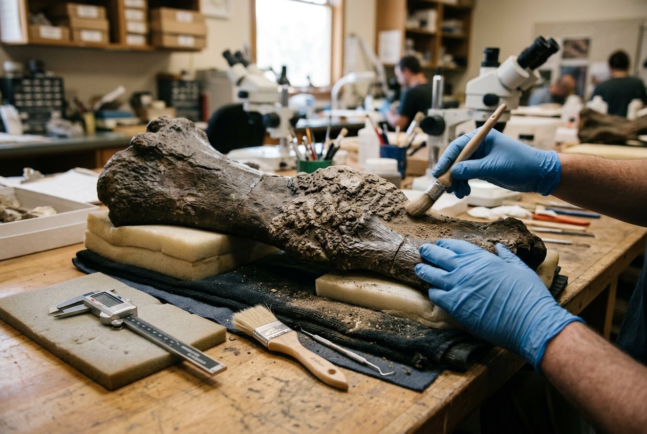
Image context: the lead image now uses an immersive fossil-preparation scene rather than a scientific composite. It fits the article because the key point is not spectacle but close material reading: healed bone, careful support, tools, and the time implied by repair.
1) Healing is the main signal
The cleanest correction is that pathology in fossils is usually about duration. A wound that kills immediately may leave little more than damage. A wound that an animal survives can leave callus, remodeling, reactive bone, fused elements, altered growth, or a distorted loading pattern somewhere else in the skeleton.[1][2][4] Those changes are what give paleopathology its leverage. They let researchers say that an extinct animal absorbed an injury into ordinary life for weeks, months, or longer.
The 2022 Scientific Reports paper on the Triceratops specimen Big John shows that principle well.[2] The famous part is the hole in the frill. The scientifically stronger part is the tissue around the hole. D'Anastasio and colleagues described plaque-like reactive bone and lytic lesions around the opening, then used histology and chemical analysis to argue that the trauma had triggered a healing response consistent with periostitis and bone repair already underway.[2] That does not merely tell us that the frill was struck. It tells us the animal kept living with the injury.
That time dimension is why fossil injuries can be so revealing without ever becoming melodrama. A healed lesion narrows the story. The animal was wounded. The wound did not end the story at once. Bone tissue responded in a specific way. Whatever behavior caused the injury, the skeleton records the aftermath rather than the cinematic instant.[2][4]
2) Diagnosis gets better when spectacle gets smaller
Pathology is also one of the places where paleontology most clearly benefits from slowing down. The Museum für Naturkunde's methods page lays out the basic logic: researchers examine external morphology macroscopically and microscopically, then study internal structure through micro-CT, synchrotron tomography, and histological thin sections.[1] In other words, a strange-looking fossil surface is only the beginning of diagnosis.
The 2020 Scientific Reports study on a Tyrannosaurus rex takes that logic further by adding what the authors call phylogenetic disease bracketing to CT-based morphology.[3] Their problem was a familiar one: abnormal bone can look dramatic without being diagnostically clear. Initial morphological assessment of the left fibula and fused caudal vertebrae narrowed the options to infection or neoplasia, but the authors then compared likely disease frequencies in close living relatives within the bird-and-reptile bracket.[3] Infection proved much more plausible than tumor, and the final diagnosis favored multiple foci of osteomyelitis.[3]
That is a valuable result partly because of what it refuses to do. It does not pretend that one glance at a damaged bone can produce certainty. It builds certainty by stages: gross form, internal structure, comparison with extant relatives, and elimination of weaker alternatives.[1][3] Paleopathology is strongest when it behaves like constrained diagnosis rather than creative storytelling.
3) A damaged skeleton does not always owe us a dramatic cause
This is where the field becomes especially useful for behavior. Fossil injuries tempt readers toward grand narratives: predator attack, ritual combat, heroic escape, brutal mating contest. Some cases may point in those directions. Many do not need that much theater. What they often show more securely is loading history and survivable disruption.
The 2024 PLOS ONE study on pathological chevrons in Plateosaurus trossingensis makes that restraint look productive.[4] Schaeffer and colleagues described pathological changes in the tail chevrons of two individuals from the Upper Triassic of southwestern Germany. The lesions required substantial force, and the authors considered several possibilities, including trampling, accidental falls, social interaction, or predatory injury.[4] But the paper's value sits less in selecting one dramatic cause than in tracing consequences. CT scans showed reactive periosteal bone and altered growth, while the authors argued that even with pathology at the distal chevrons, the tail could likely continue functioning as a muscle attachment site without catastrophic loss of performance.[4]
That is a strong paleobiological result. A skeleton can preserve that an injury happened low on the tail, that new bone formed, that growth was disrupted, and that the animal survived without the whole structure becoming useless.[4] Those are already meaningful facts about mechanics and life history. The field gets stronger when it accepts how much can be said before the exact cause is pinned down.
4) Disease belongs in dinosaur biographies too
Trauma tends to dominate public imagination, but pathology is broader than trauma. The hadrosaur vertebrae described in the 2020 Scientific Reports paper on Langerhans Cell Histiocytosis are a useful reminder that fossil disease can include tumor-like disorders as well.[5] The authors compared hadrosaur vertebral lesions with human cases diagnosed in life and found the fossil material consistent with LCH, a benign osteolytic tumor-like condition that commonly affects the skeleton.[5]
What matters in that paper is not just the label. It is the claim that disease diversity in extinct animals has probably been under-read because diagnosis is difficult and isolated lesions are easy to misclassify.[5] Once a case is constrained well enough, pathology stops being an embarrassment to the idealized dinosaur body and starts becoming part of ordinary vertebrate life. Dinosaurs healed, inflamed, fused, deformed, and sometimes developed lesions that fit recognizable disease processes.[1][3][5]
That widens the interpretive field. Paleopathology can inform behavior, but it also bears on immune response, growth, tissue turnover, and the evolutionary depth of disease categories that humans tend to imagine as modern.[1][5] A pathological fossil is not merely a damaged specimen. It is a reminder that extinct animals inhabited the same biological reality of stress, repair, and vulnerability as living ones.
5) What fossil pathology does best
The strongest paleopathology therefore sits in a narrow but rich zone. It can show that an injury healed, that infection reached bone, that growth was rerouted, that motion or loading changed, or that a lesion fits one diagnosis better than another when modern comparisons and imaging are brought to bear.[1][2][3][4][5] It can sometimes support inferences about combat, feeding accidents, predation, social interaction, or locomotor strain. It can also put hard limits on overreach.
That last point is the reason the field matters. Fossils rarely preserve the whole event. They preserve the biological residue of the event. A frill lesion with reactive bone tells us more securely that Triceratops survived trauma than why the blow landed.[2] A pathological tyrannosaur fibula can support osteomyelitis more strongly than a dramatic one-line story about how the infection began.[3] A distorted Plateosaurus tail can register force and recovery without naming the exact accident.[4]
Read that way, pathologic bone is one of paleontology's most intimate archives. It records an extinct animal under stress, then records what happened next. The value lies less in prehistoric violence than in survivorship written into tissue. Deep time becomes legible here because bone kept a memory of living through damage instead of merely receiving it.
Sources
- Museum für Naturkunde Berlin, "Paläopathologie fossiler Wirbeltiere" - overview of paleopathology as a research field, including questions about disease history, bone healing, behavior, and methods such as micro-CT, synchrotron tomography, and histology.
- Ruggero D'Anastasio, Jacopo Cilli, Flavio Bacchia, Federico Fanti, Giacomo Gobbo, and Luigi Capasso, "Histological and chemical diagnosis of a combat lesion in Triceratops," Scientific Reports 12 (2022).
- C. A. Hamm and colleagues, "A comprehensive diagnostic approach combining phylogenetic disease bracketing and CT imaging reveals osteomyelitis in a Tyrannosaurus rex," Scientific Reports 10 (2020).
- J. Schaeffer, E. Wolff, F. Witzmann, G. S. Ferreira, R. R. Schoch, and E. Mujal, "Paleobiological implications of chevron pathology in the sauropodomorph Plateosaurus trossingensis from the Upper Triassic of SW Germany," PLOS ONE 19, no. 7 (2024).
- Bruce M. Rothschild, Darren Tanke, Frank Rühli, Ariel Pokhojaev, and Hila May, "Suggested Case of Langerhans Cell Histiocytosis in a Cretaceous dinosaur," Scientific Reports 10 (2020).