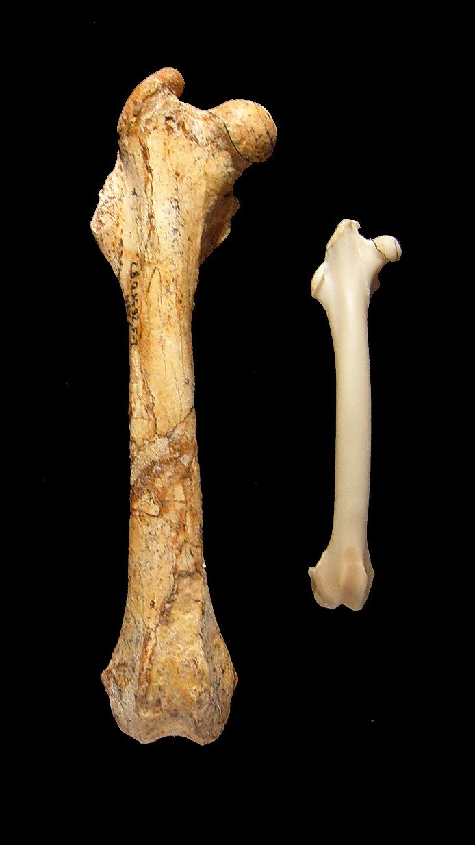
One femur is thick enough to make the other look like a splinter. In the cover photograph, the uploader identifies the larger bone as Nuralagus rex, the extinct giant rabbit of Menorca, and the smaller as a living European rabbit. The comparison delivers scale in an instant. It cannot show how long either animal took to build that scale.
That second question sits inside the bone. In 2023, researchers cut and examined a growth series of Nuralagus femora from Pliocene fissure deposits at Punta Nati. The site is probably early Pliocene, although its age is not directly radiometrically fixed. The team was testing an idea more demanding than “islands produce giants.” A larger mammal normally grows and matures more slowly than a smaller one, so the rabbit’s size alone could explain some delay. The real test was whether its development remained unusually slow after body mass and ancestry were taken into account.[1][2]
It did. The fitted rate at which Nuralagus approached adult femoral area was almost five times lower than in the directly comparable European brown-hare sample. The models placed sexual maturity several years later than body-mass scaling predicted, while the timing of femoral joint fusion independently pointed in the same direction.[1] Menorca’s giant rabbit was not a continental rabbit enlarged at the usual pace. It stretched growth across a radically longer schedule.
Image context: the cover is a real, user-contributed photograph of whole femora, not a reconstruction or a microscopic plate. Its identifications come from the uploader and no repository or scale is supplied. The smaller bone is labeled as the European rabbit Oryctolagus cuniculus; the living calibration in the study came instead from known-age European brown hares, Lepus europaeus. The photograph illustrates gross contrast; the separately documented research sample establishes the clock.[1][6]
The animal was strange before anyone cut the bone
Nuralagus rex lived on Menorca around the opening of the Pliocene and is the largest wild leporid yet known. Even its mass needs a methodological footnote. The 2011 description estimated an average near 12 kilograms; a later set of lagomorph-specific regressions produced an estimate around 8 kilograms after rejecting measurements that did not scale reliably or were distorted by specialized anatomy.[2][3] Both estimates describe a rabbit far outside the usual wild range. Neither is a weight read directly from a fossil.
Its skeleton was not simply supersized. The vertebral column was short and comparatively stiff, limiting the flexion and extension that power a rabbit’s bounding gait. The hands and feet were short, with splayed digits; the limbs were robust; the palms bore weight. Reduced eye sockets, auditory bullae, and braincase proportions suggested less investment in the sensory vigilance typical of prey under constant pursuit. The original authors read this package as low-gear locomotion and energy economy in an island ecosystem with few mammalian competitors and no known large terrestrial predator.[2]
Those are anatomical and ecological inferences. The bones do not preserve a rabbit moving, listening, or reproducing. Histology offered a different kind of evidence: not another reconstruction of what the animal looked like, but a record of how its skeleton accumulated.
A femur is a structure and an archive of construction
Bone is not poured once and left unchanged. Growing mammals deposit tissues at different rates, open vascular canals as new bone forms, pause or slow deposition seasonally, and remodel older cortex from within. Under a microscope, that activity can leave changes in tissue organization and lines of arrested growth, often shortened to LAGs. A dense outer zone with little vascularity can signal that rapid body growth is nearing its end.[1][4]
The principle is powerful and easy to oversimplify. A line is not automatically one birthday. Remodeling can erase early tissue. A season can produce several closely packed marks, while another mark may be difficult to trace around the full shaft. The distance between rings is also deceptive: adding the same thickness to a wider bone produces more new cross-sectional area than adding it to a narrow one.[1][4]
This is why the Nuralagus study did not begin by counting dark bands and declaring ages. It first built a calendar in a close living relative whose life events were known.
The difficult work began in red stone
The Punta Nati bones were locked in hard, carbonate-cemented red silt inside karst fissures. Preparators protected exposed fossils, immersed blocks in a 10 percent acetic-acid solution for two days, rinsed them in fresh water for two more, dried and reconsolidated them, and repeated the cycle. Completely dissolving the surrounding rock took about four years.[1]
Twelve measurable Nuralagus femora ultimately entered the growth analysis. From the middle of each shaft, the team removed a block roughly two centimetres long, embedded it in epoxy, cut it with a diamond saw, fixed it to glass, and ground it into a thin section for microscopy. Histology extracts information by sacrificing a small part of the specimen. That cost helps explain why fossil samples remain narrow even when the questions are broad.[1]
The researchers traced each annual growth line on micrographs and measured the entire cross-sectional area enclosed by it, including the medullary cavity. Area, rather than a single radial distance between lines, became the size variable in the growth curves. The choice sounds technical; it prevents the widening geometry of a femur from masquerading as a biological slowdown.[1]
A living hare supplied the calendar
The comparison sample of European brown hares included animals of known age and sex. Some received fluorescent bone labels on a controlled schedule, giving the researchers dated marks against which natural tissue transitions could be read. An additional known-age group carried ordinary winter growth lines and extended the sequence beyond maturity.[1]
The labeled bones connected texture to events. Fast-growing fibrolamellar tissue dominated before weaning. After weaning, slower lamellar tissue with primary osteons appeared. Around puberty, the cortex shifted again toward more slowly deposited lamellar bone; near sexual maturity, growth became extremely slow and the outer cortex formed an external fundamental system. In the brown-hare growth models, reaching 99 percent of asymptotic size corresponded reasonably well with independently observed maturity, making that point a defensible proxy rather than an arbitrary percentage.[1]
This calibration is the hinge of the method. A fossil growth curve has no meaning by itself. It becomes biologically legible only after a living comparison links tissue, age, and reproductive timing—and even then, the comparison supplies an inference, not a time machine.
The giant rabbit was already growing slowly in its first year
The contrast appeared deep in the cortex. Before weaning, Nuralagus deposited mainly lamellar bone with primary osteons, a slower tissue than the hare’s early fibrolamellar complex. A weaning-related band formed before the first winter line, showing that the young rabbit had left milk during its birth year. From there, vascular canals became progressively scarcer. After the first winter they were sparse; by the end of the third year, the outer tissue was essentially without them.[1]
Its annual lines crowded together so closely that simple visual spacing could make the onset of skeletal maturity ambiguous. The cross-sectional area measurements and fitted growth curves did the heavier work. They separated the sample into two size groups. The authors interpreted the larger animals, which approached adult size earlier, as females and the smaller, later-maturing animals as males—a plausible assignment because female leporids are commonly the larger sex, but still an assignment made from growth pattern rather than preserved reproductive anatomy.[1]
The model estimated maturity at about 3.6 years for the larger group and 6.2 years for the smaller group. A body-mass model spanning living herbivorous mammals predicted roughly 1.28 years for an animal of Nuralagus’ size. Its fitted rate of approach to asymptotic femoral area was almost five times lower than in the comparable brown-hare sample. That coefficient describes the growth curve; it is not a direct measurement of tissue deposited per day.[1]
One model did not carry the conclusion alone. At the upper end of the femur, joint surfaces in Nuralagus began fusing years later than the equivalent surfaces in hares. At the lower end, the suture was still visible at an inferred age of about four years, a state comparable to a hare near maturity. The sequence of epiphyseal fusion therefore supported the late timetable recovered from the growth curves.[1]
Large bodies did not explain the delay away
Calling this “island gigantism” is a description of direction, not a mechanism. Bigger bodies usually take longer to build. To ask whether Menorca changed the pace of life as well as body size, the study compared maturity and mass across 81 living herbivorous mammal taxa while accounting for shared ancestry. Nuralagus remained an extreme late-maturing outlier, especially beside living leporids.[1]
That result favors the island-syndrome interpretation: low external mortality can reward delayed reproduction, prolonged growth, and survival rather than the fast turnover useful where predators remove many young animals. Scarce or seasonal resources can also favor economical movement and slow tissue deposition. A giant need not reach its size through a growth spurt; it can get there by growing gently for much longer.[1]
The pattern now extends beyond one rabbit. A 2024 analysis of metabolic data from 2,813 living tetrapod species found that island mammals and birds tended toward lower mass-adjusted metabolic rates and longer generation times than non-island endotherms.[5] That broad result gives the fossil interpretation comparative context. It does not prove which pressure—predator release, competition, resource limits, or their interaction—set Nuralagus on its particular path.
The clean edge of the evidence
The case is strong because different observations converge: slowly deposited and weakly vascularized cortex, annual area increments, fitted growth trajectories, late epiphyseal fusion, low-gear gross anatomy, and a phylogenetically corrected comparison. It is bounded because the fossil sample contains only 12 measurable femora from one insular taxon.[1]
No section records an actual mating or birth. The proposed sexes are inferred. Annual marks can be locally hard to follow, and the onset of the outer slow-growth system is subtle precisely because the animal was already depositing bone at a low rate. Body-mass estimates have shifted with the choice of regression. Histological sampling is destructive, limiting replication. The authors explicitly call for equivalent data from other island giants before treating the result as universal.[1][3]
That boundary does not reduce the femur to a curiosity. It shows what a good anatomical method can do. The whole bone says that Nuralagus was large and heavily built. Its articulations say that development remained unfinished for years. Its microscopic fabric measures a sustained retreat from speed. The comparative model then asks the decisive question: was the delay merely what size predicts? The answer is no.
The smaller femur in the photograph makes the giant easy to see. The more remarkable difference is invisible there. Menorca’s rabbit reached enormity not by rushing through growth, but by keeping youth open for years.
Sources
- Meike Köhler and colleagues, “Insular giant leporid matured later than predicted by scaling,” iScience 26 (2023) — institutional open-access copy of the primary study of Nuralagus femoral histology, living-hare calibration, growth models, maturity estimates, epiphyseal fusion, and stated limitations.
- Josep Quintana, Meike Köhler, and Salvador Moyà-Solà, “Nuralagus rex, gen. et sp. nov., an endemic insular giant rabbit from the Neogene of Minorca,” Journal of Vertebrate Paleontology 31 (2011) — original description of the taxon, skeleton, locality, locomotor anatomy, sensory reductions, and initial mass estimate.
- Blanca Moncunill-Solé and colleagues, “The weight of fossil leporids and ochotonids: body mass estimation models for the order Lagomorpha,” Journal of Zoology 295 (2015) — UAB institutional record for the primary study of lagomorph-specific regressions and the approximately 8-kilogram Nuralagus estimate.
- Christian Kolb and colleagues, “Mammalian bone palaeohistology: a survey and new data with emphasis on island forms,” PeerJ 3 (2015) — open review of mammalian bone tissues, vascularity, growth marks, and the interpretive scope of palaeohistology.
- Ying Xiong and colleagues, “Convergent evolution toward a slow pace of life predisposes insular endotherms to anthropogenic extinctions,” Science Advances 10 (2024) — broad comparative study of metabolism, generation length, and insularity across living tetrapods.
- Wikimedia Commons, “Nuralagus rex & rabbit femur bone” — source page and provenance record for the public-domain comparison photograph used as the article image.