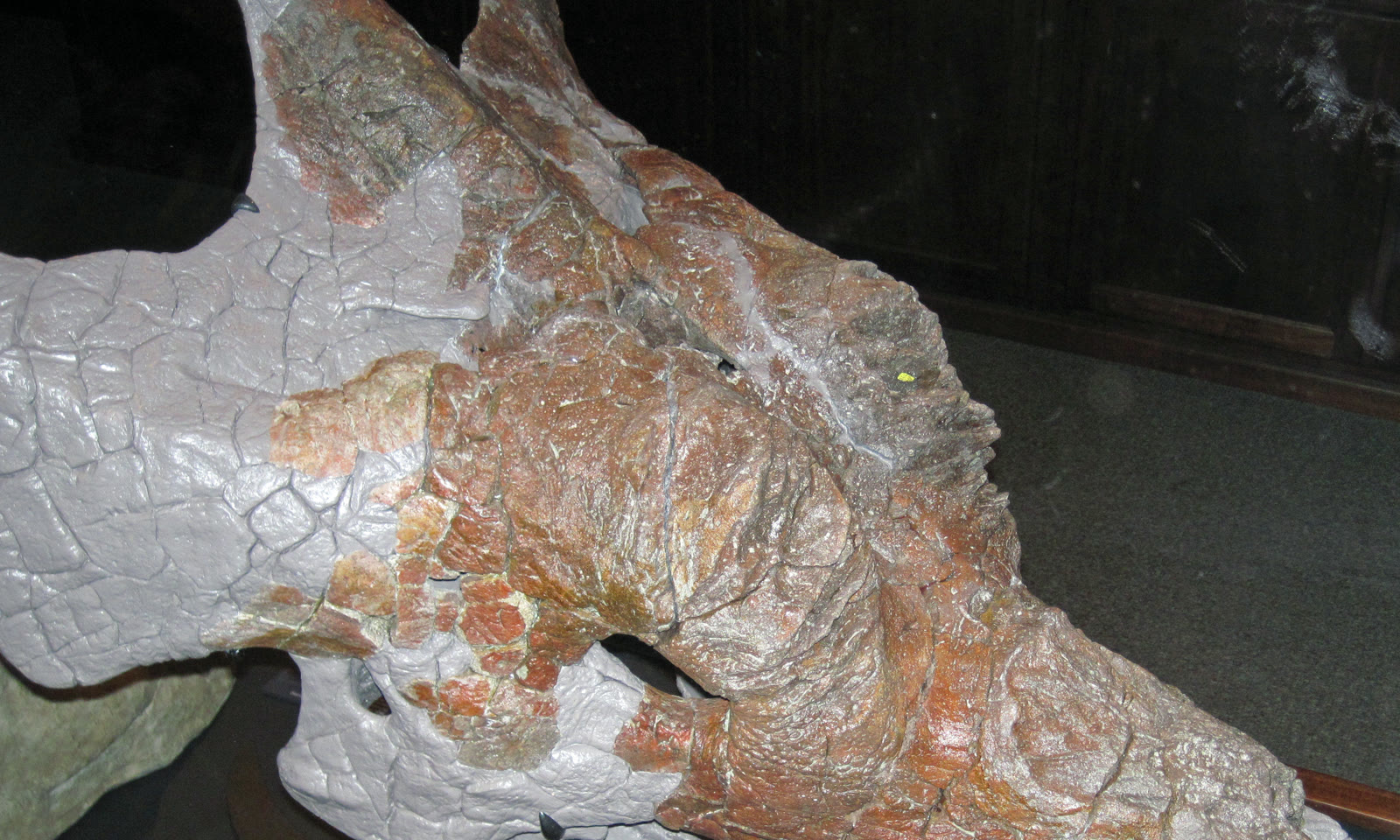
The face in the cover photograph has no face.
It is the partial skull of Achelousaurus horneri, a horned dinosaur from the Late Cretaceous of Montana. Across the snout and above the eye, projecting horn cores have given way to blunt, rough bosses. The fossil preserves those bony surfaces in exquisite detail. It does not preserve the tissue that met them, the contour that tissue made in life, or its color.[3][6]
That absence is the central problem of reconstructing extinct animals. Skin, muscle, blood vessels, glands, and air sacs usually vanish, yet a skeleton without them is not an animal. Adding them back by intuition merely hides the gap. Paleontology needs a way to ask which missing structures are strongly supported, which require an evolutionary change, and which remain plausible but untested.
The extant phylogenetic bracket supplies that discipline. Developed most fully by anatomist Lawrence Witmer, it uses the closest living relatives on either side of an extinct group and the physical traces soft tissues leave on their bones. For non-avian dinosaurs, the two principal witnesses are birds and crocodilians: the surviving branches that frame them within Archosauria.[1] Neither witness is a dinosaur costume. Together they form a controlled comparison.
Image context: the cover is a real photograph of the Achelousaurus horneri holotype, MOR 485, at the Museum of the Rockies—not a life restoration.[6] The rough bosses are especially apt because the study that interpreted centrosaurine facial tissues examined this anatomy directly.[3] Cropping brings the bone surface closer; it does not supply the missing skin.
Bone remembers contact
Bone is living tissue during an animal’s life. Muscles pull on it. Tendons anchor into it. Blood vessels cross and enter it. Horn sheaths and other skin structures grow against it. Those relationships can produce ridges, pits, grooves, canals, altered surface textures, or microscopic fibers.
Such a feature becomes an osteological correlate when observation in a living animal establishes a causal association between a soft tissue and a bony mark.[1] The distinction matters. A groove does not mean “blood vessel” because it looks vaguely tube-like. It supports that reading when anatomy shows vessels repeatedly occupying comparable grooves. The fossil mark and the living relationship make a testable pair.
This is why reconstruction begins at the contact surface rather than at the silhouette. Witmer’s framework asks researchers to identify a soft tissue in living relatives, document whether it has a consistent skeletal correlate, and then look for that correlate in the fossil.[1] The method does not make decay reversible. It makes the reasoning inspectable.
A broad reconstruction can be built from many such local tests. In a study of sauropodomorph forelimbs, Alejandro Otero synthesized dissections and anatomical data from living birds and crocodilians, mapped attachment evidence on fossil bones, and evaluated the musculature one structure at a time.[2] The result was not a single guess called “the arm.” It was an inventory of separate claims, each carrying its own anatomical support.
Two sides of the bracket set the price of an inference
If a tissue and its bony correlate occur in both birds and crocodilians, the simplest explanation is that their common archosaur ancestor possessed the relationship. Finding the predicted mark in a non-avian dinosaur then makes the reconstruction especially well supported. Rejecting it would require the association to disappear along the dinosaur branch despite the fossil evidence.[1]
When only one living side supports the relationship, the inference is weaker, not forbidden. A gain or loss somewhere in the family tree must be proposed. When neither side supplies a homolog, a novel structure may still have existed, but the bracket alone cannot carry the claim. Witmer’s levels rank this escalating inferential cost.[1] They are not percentages, and they do not turn biology into a traffic-light system. Their value is that they expose where additional assumptions enter.
That hierarchy also prevents two common mistakes. The first is to copy everything from the closest-looking living animal. A crocodile’s jaw muscle may be informative because of homology and matching bone contacts; a rhinoceros horn is not automatically informative because a dinosaur boss happens to look rough. The second mistake is to treat uncertainty as failure. A lower-level inference can be a productive hypothesis if it is labeled honestly and tested with broader evidence.
The boss where the bracket goes quiet
The Achelousaurus bosses make a revealing boundary case. Ceratopsid horn cores and bosses have no direct homologs in living birds or crocodilians, so Hieronymus and colleagues classified their soft-tissue reconstruction as a Level III inference.[3] The bracket identifies the evidential gap; it does not fill it.
The researchers therefore widened the comparison without pretending that analogy had become ancestry. They sampled 96 specimens representing 84 living amniote taxa, pairing known skin structures with the bone beneath them.[3] Rhinoceroses, muskoxen, bovids, birds, and reptiles became experimental references for how different kinds of scales, sheaths, pads, and horns modify a skull. Surface morphology and, where available, histology helped distinguish superficially similar arrangements.
That comparison argued against simply erecting a tall rhinoceros-like horn over every rough boss. On adult Achelousaurus, the pitting, grooves, and bony “fins” more closely matched attachment for a thick pad of cornified epidermis. The orientation of those features even suggested how the covering grew across the bone. The authors interpreted that pad as an evolutionary transformation of the horn sheath found in earlier centrosaurines.[3]
This is a constrained reconstruction, not a recovered portrait. “Thick cornified pad” says something substantial about tissue class and attachment. It does not deliver the pad’s precise outer contour, wear pattern, or color. The bone is a witness, not a mold.
Direct preservation can answer back
Occasionally, exceptional fossils preserve the missing tissue itself. These specimens do not make comparative inference obsolete; they test it.
The ceratopsian Psittacosaurus offers a particularly useful example. Specimen SMF R 4970 preserves polygonal scales on the ventral surface of the cheek horn and a displaced dark patch of soft tissue that Bell and colleagues interpreted as a dorsal keratinous covering. They judged that covering more comparable to a fingernail than to a complete horn sheath. Crucially, this distribution agreed with differences in the underlying bone texture: the smoother, porous ventral surface did not fit a thick keratinous covering.[5]
Exceptional preservation therefore sharpened the soft-tissue boundary rather than merely confirming that “there was keratin.” It also warns against transplanting the exact arrangement to another ceratopsian. Psittacosaurus is not Achelousaurus. Its preserved skin is a calibration point for how bone and covering correspond, not a universal face supplied by a better fossil.
Silence in the skeleton is not absence in life
Positive evidence has limits, and negative evidence has sharper ones. A tissue can be present without leaving an obvious, preserved mark.
Petermann and Sander tested this problem on living rabbit, alligator, and turkey femora. They first dissected and mapped the actual muscle attachments, then searched thin sections for microscopic indicators such as Sharpey’s fibers, vascular orientation, and frayed bone margins. Only about 60 percent of the mapped attachment sites were detected histologically. Features that looked suggestive of attachment also appeared outside the mapped sites.[4]
Even destructive access to modern bone, backed by a dissection map, produced false negatives and possible false positives. A fossil offers less: a limited surface, a tiny number of permissible sections, and a history of burial, breakage, preparation, and remodeling. Smoothness cannot by itself prove that no muscle or other tissue was present.
The asymmetry is important. A well-validated correlate in the predicted place can support a specific reconstruction. Failure to find a correlate may leave several explanations open. Good skeletal anatomy therefore does not maximize the amount of flesh inferred; it separates evidence of presence from mere absence of evidence.
Behavior sits farther down the chain
Soft-tissue anatomy is already an inference. Behavior usually adds another sequence: a muscle was present, its size and path are reconstructed, its leverage is estimated, a movement becomes possible, and that possibility is translated into an act.
The same distance applies to the Achelousaurus bosses. A cornified pad makes repeated contact more mechanically plausible than delicate bare skin. It does not preserve a clash, a shove, courtship, species recognition, or any other single episode. Hieronymus and colleagues tested living analogues for associations between structure and behavior, but they also stressed that an analogy extends only as far as the relationship demonstrated in the living system.[3]
That is the deeper virtue of the bracket. It does not force every extinct animal into a cautious gray blur. It lets strong details remain strong while preventing them from underwriting an entire scene. A dinosaur can have a well-supported set of muscles, a defensible facial covering, and behavior that remains open.
Birds and crocodilians cannot tell us everything that once occupied a dinosaur skeleton. They can show which bone-and-tissue relationships survived on both sides of the family tree. Fossils can preserve the predicted consequences, contradict them, or reveal novelties for which no living witness exists. Exceptional skin can check the result. Every layer changes the confidence of the next.
Reconstruction becomes most vivid when those labels stay attached. The finished animal is not hidden intact inside the stone. It is assembled claim by claim from what bone remembers, what living bodies demonstrate, and what the evidence still refuses to say.
Sources
- Lawrence M. Witmer, “The Extant Phylogenetic Bracket and the Importance of Reconstructing Soft Tissues in Fossils” (1995) — the foundational statement of phylogenetically constrained soft-tissue inference and its levels of support.
- Alejandro Otero, “Forelimb musculature and osteological correlates in Sauropodomorpha (Dinosauria, Saurischia),” PLOS ONE 13 (2018) — a muscle-by-muscle application of living archosaur comparisons to fossil forelimbs.
- Tobin L. Hieronymus, Lawrence M. Witmer, Darren H. Tanke, and Philip J. Currie, “The Facial Integument of Centrosaurine Ceratopsids: Morphological and Histological Correlates of Novel Skin Structures,” The Anatomical Record 292 (2009) — comparative evidence for cornified pads over the bosses of Achelousaurus and Pachyrhinosaurus.
- Holger Petermann and P. Martin Sander, “Histological evidence for muscle insertion in extant amniote femora: implications for muscle reconstruction in fossils,” Journal of Anatomy 222 (2013) — an experimental measure of false negatives and ambiguous signals in muscle-attachment reconstruction.
- Phil R. Bell et al., “The exquisitely preserved integument of Psittacosaurus and the scaly skin of ceratopsian dinosaurs,” Communications Biology 5 (2022) — direct skin preservation used to test the relationship between a cheek horn and its covering.
- Wikimedia Commons, “File:Achelousaurus bosses side.jpg” — source and license page for James St. John’s photograph of the holotype skull MOR 485 used as the cover image.