The crab came through beautifully. The biggest dinosaur bones did not. In a 2015 account of experiments at Australia's nuclear science agency, ANSTO, Ada Klinkhamer of the Australian Age of Dinosaurs Museum described seeing crustacean fossils inside their enclosing rock with neutron tomography. Some large sauropod specimens, however, were too thick and dense for the beam to penetrate their centres successfully.[1]
That uneven result is a useful introduction to fossil scanning. An instrument does not simply make stone transparent. It needs enough radiation to pass through the specimen, and enough difference between its materials to tell them apart. Neutron imaging offers another way to obtain that difference. Its value lies in what a particular fossil lets it see.
A different kind of shadow
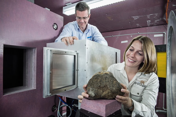
At ANSTO's Dingo instrument, a sample rotates while a detector records a succession of views. A computer combines these projections into a three-dimensional volume, allowing researchers to inspect virtual slices through the object. The broad procedure resembles an X-ray CT examination; the materials that stand out can be quite different.[1][2]
ANSTO describes neutron radiography as particularly useful for seeing hydrogen-bearing substances inside metal or ceramic objects. Water or oil can become conspicuous where another imaging method would emphasize the container. The same principle gives paleontologists a second opportunity when a fossil and its surrounding rock are difficult to distinguish with X-rays.[2]
Two requirements therefore have to be separated. Penetration gets a usable beam through the object. Contrast makes adjacent materials distinguishable in the resulting image. Passing the first test does not guarantee the second. Nor does a finer grid of image pixels automatically supply a missing boundary: researchers need a signal that actually changes across it.
The tooth inside the grey patch
A 2020 study led by Clément Zanolli compared X-ray and neutron scans of fossil primate jaws and teeth. One Pleistocene molar from Java, specimen SMF-8888, looked internally almost uniform in its X-ray image. Neutrons distinguished enamel from dentine and revealed projections of the pulp cavity that were barely discernible before.[3]
Another Javanese tooth, Sangiran 7–65, exposed a more troublesome ambiguity. Bright fringes in synchrotron images could be mistaken for the enamel's inner boundary. The neutron scan placed that boundary closer to the chewing surface. At one illustrated position, the misleading reading suggested 2.08 millimetres of enamel; the neutron result indicated at most 1.15 millimetres.[3]
A boundary error becomes a biological error as soon as someone measures it. Enamel thickness is an anatomical property; a bright edge produced by imaging is not. Comparing methods gives researchers a way to question an apparently persuasive shape before turning it into a claim about an extinct animal.
Keep the surrounding rock in the story
The target can also be the deposit around the fossil. In 2021, Holly Smith and colleagues examined thirteen blocks from three Pleistocene caves in Sumatra. These cemented mixtures of sediment, rock fragments and animal remains are called breccias. Synchrotron X-ray attempts had failed because the samples attenuated the beam too strongly.[4]
Neutrons introduced their own obstacle. Early scans lasting 10–24 hours produced poor results or none because the samples contained too much moisture. Drying improved the results, although some blocks remained difficult to read. The team's eventual analysis supported several depositional episodes involving water and sediment gravity flows, rather than treating the enclosed remains as a simple, undisturbed assemblage.[4]
Here, preparation has an unusual goal: retain the arrangement that digging out an attractive tooth might erase. A loose fossil can be identified and measured, but the positions of nearby fragments and empty spaces help explain how its burial happened. The surrounding material is part of the evidence. Imaging can make it worth keeping.
The specimen brings its later history, too
Hydrogen sensitivity can also reveal a museum's handiwork. Zanolli's team found bright areas where glue had consolidated a broken fossil jaw; the corresponding X-ray view did not show the same information.[3] A conspicuous feature in a scan may belong to a repair made millions of years after burial. Preparation records matter alongside geological ones.
There is also a practical consequence after exposure. Neutron irradiation can leave specimens temporarily radioactive. In the Sumatran study, the rapid scanning approach allowed the samples to be cleared and returned within a week.[4] That is a reported outcome for those samples and conditions, not a universal collection-return timetable.
The failed sauropod scans remain a useful companion to these successes.[1] They stop the technique from becoming a promise that every sealed object will yield its contents. A persuasive fossil image depends on matching the beam to the material, recognizing what preparation has changed, and checking which boundaries the data actually support. The reward can be an inner tooth surface or a history of cave sedimentation—evidence recovered while the object still holds together.
Sources
- ANSTO, with an account by Ada Klinkhamer, “Fossils go nuclear” (2015) — early Dingo trials, crustacean and large-bone scans, and photograph.
- ANSTO, “Dingo — Neutron Imaging” — instrument overview, tomography and contrast between materials.
- Clément Zanolli and colleagues, “When X-Rays Do Not Work,” Frontiers in Ecology and Evolution (2020) — comparisons of fossil dental tissues and the visibility of conservation materials.
- Holly E. Smith and colleagues, “High-resolution high-throughput thermal neutron tomographic imaging of fossiliferous cave breccias from Sumatra,” Scientific Reports (2021) — sample moisture, depositional interpretation and post-scan clearance.