Leanchoilia superlata looks almost legible too quickly. The body is small, arthropod-like, and divided into a head, trunk, limbs, and tail region. Then the front end complicates everything. Three long flagella extend from each of the great appendages, making the animal seem as if it carried delicate antennae, fishing lines, or ornamental feelers in front of the body.[1][2] The best reading starts by refusing those quick metaphors. In Leanchoilia, the great appendages are not decoration. They are the method problem.
The animal belongs to the Cambrian Burgess Shale record, where soft-bodied arthropods were flattened into fine-grained mud in ways that preserve outlines, carbon films, and sometimes internal traces that ordinary fossilization would erase.[1][5] That exceptional preservation gives the fossil power, but it also creates a trap. A flattened animal can look simpler than it was. A long appendage trace can be sensory, grasping, damaged, displaced, or a mix of original anatomy and burial geometry. Leanchoilia matters because several evidence layers have to be read together: the great appendages, the paired eyes, the trunk limbs, the telson, and the gut.[2][3][4][5]
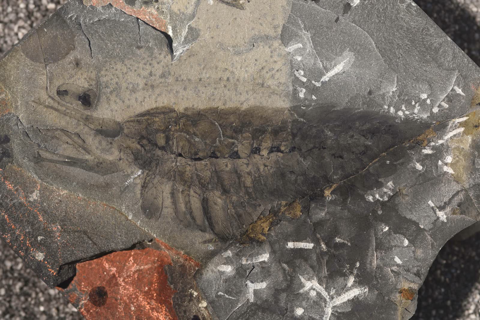
The cover image shows specimen USNM PAL 83943b, photographed through Smithsonian material and hosted on Wikimedia Commons.[6] It is the right visual anchor because it keeps the article inside the actual fossil record. The photograph does not hand over a polished life pose. It shows a compressed Burgess animal whose scientific force comes from repeated anatomical reading: where the appendages begin, how the body axis runs, what belongs to the trunk, and why the long traces at the front are too important to be treated as visual flourish.[6]
The Great Appendage Is Not One Tool
The Royal Ontario Museum's Burgess Shale profile places Leanchoilia superlata among megacheiran arthropods, a group whose name points to the conspicuous "great appendage" at the front of the body.[1] That label can make the part sound single-purpose, as if one enlarged limb solves the animal. The more useful question is how that appendage was partitioned.
Haug, Briggs, and Haug's 2012 redescription is valuable because it treats Leanchoilia as an anatomical matrix rather than as a silhouette.[2] They break the animal into describable units and use that method to test possible function. The great appendage includes a proximal portion and long flagellar structures, so its job cannot be reduced to a claw. It may have combined tactile exploration, prey detection, and food-handling roles.[2] In other words, the appendage was probably a front-end interface between the animal and its immediate environment.
That matters because Cambrian "great appendage" animals often get folded into a predator image too quickly. Raptorial front limbs are real evidence in several early arthropods, but not every long anterior structure is a spear. In Leanchoilia, the long flagella make a narrow, more interesting claim possible: the animal may have sampled the space in front of it while the rest of the body remained close enough to the seafloor for benthic or near-bottom life to matter.[1][2] A sensor array is less cinematic than a weapon. It is also more faithful to the anatomy.
The safe interpretation is therefore plural. The great appendages probably helped Leanchoilia find, touch, or manipulate things. They may also have helped bring food toward the mouth. But the evidence does not require a single dramatic feeding stroke. It asks for a working front end.
Eyes Change The Reading Order
The front of Leanchoilia becomes sharper once the eyes enter the frame. Garcia-Bellido and Collins's reassessment of the Burgess Shale genus identified new characters in L. superlata, including two pairs of eyes and a dorsal double carina bracketing the body axis.[3] The eyes are not trivia. They change how the appendages should be imagined.
If an animal has long anterior tactile structures and paired visual organs, the front end is doing more than passively sweeping sediment. It is coordinating sensory input. Schoenemann and Clarkson's study of Leanchoilia eyes argued that the visual system was likely adapted to relatively low light, with the animal probably crepuscular in habit.[4] That inference should be handled carefully. It does not let us reconstruct a daily schedule with confidence. It does, however, make the animal less like a blind bottom rake and more like a visually equipped Cambrian arthropod operating in a light environment that mattered.[4]
This is one reason an anatomy-and-method reading works better than a simple species profile. The eyes and appendages constrain each other. If the flagella were sensory, the eyes give a second sensory channel. If the animal was active near the substrate, the eyes and appendages together could help it navigate a dim, cluttered, particle-rich environment. A single fossil part can suggest a story; the paired systems make the story harder to dismiss.[2][4]
The boundary remains important. Compound-eye interpretations depend on preservation quality, comparisons with living arthropod visual systems, and whether the repeated structures in the fossil are genuinely ommatidial or lens-like rather than decay textures.[4] Leanchoilia is useful because it makes that discipline visible. The fossil does not merely say "Cambrian arthropods had eyes." It asks what kind of eye evidence can survive and how far functional inference should go.
The Gut Is A Preservation Test
The most methodologically demanding part of Leanchoilia is not the long appendage. It is the gut. Nicholas Butterfield's 2002 paper on Leanchoilia guts addressed three-dimensional structures in Burgess Shale-type fossils, asking how internal features could be preserved and interpreted when the whole animal has otherwise been compressed into a slab.[5] That paper matters because it moves the article away from surface anatomy and into taphonomy.
A gut trace is powerful because it can reveal biology that the outline hides: where food passed, how internal space was organized, and whether dark or three-dimensional features belong to anatomy rather than mineral accident.[5] But gut traces are also dangerous evidence. Internal structures can be distorted, replaced, compacted, or confused with other decay products. Butterfield's contribution is not simply that Leanchoilia had preservable guts. It is that three-dimensional Burgess structures need an interpretive rulebook.[5]
That rulebook changes the way the whole animal reads. The great appendages suggest how the animal met the world. The eyes suggest how it sensed the world. The gut asks what happened after capture, manipulation, or ingestion. Once all three are present, Leanchoilia stops being a Cambrian outline with spectacular feelers. It becomes a body system: detect, orient, handle, ingest, digest, preserve.
This sequence is exactly where paleontology can overreach if it is not careful. A gut does not identify prey by itself. A raptorial or tactile appendage does not prove a specific hunting style. Low-light eye function does not prove nocturnal behavior in the modern sense. The stronger claim is integrated and bounded: Leanchoilia preserves enough anatomy to discuss an active animal with coordinated sensory, feeding, and internal systems, while still leaving exact behavior inferred rather than observed.[2][4][5]
Why The Body Plan Resists A Modern Shortcut
The temptation is to call Leanchoilia shrimp-like, spider-like, or vaguely crustacean, then move on. That is usually a sign that the fossil has been made too comfortable. ROM's profile and the genus reassessment both keep the animal inside a more technical early arthropod problem rather than a modern category.[1][3] The great appendage links it to megacheiran debates. The body segments and appendages place it among early euarthropod-grade animals. The eyes and gut make it biologically vivid without turning it into a living analog.
This distinction matters because Cambrian fossils are often explained through resemblance first. Resemblance is useful for entry. It is weak as a conclusion. A modern shrimp has a crown-group crustacean body plan with its own specialized mouthparts, carapace, and life history. Leanchoilia is older, stranger, and less settled. Its anterior appendages are not antennae in the ordinary modern crustacean sense. Its eyes are not a license to make it behave like a crab, mantis shrimp, or spider. Its gut is not a perfect window into every meal.[1][2][5]
The better comparison is structural. Leanchoilia shows that early arthropods were already building complex front ends, not merely adding legs to worm-like bodies. It also shows that the Cambrian record can preserve multiple body systems in one animal often enough to make function discussable. That is the real payoff. The animal does not need to be a direct ancestor or a perfect modern parallel. It is valuable because it captures a working solution from a time when arthropod head organization, sensory systems, and feeding appendages were still being sorted across many forms.[2][3]
What The Fossil Still Teaches
The strongest current reading of Leanchoilia is neither monster story nor tidy ancestor story. It is a lesson in how to assemble an animal from partial but unusually rich evidence. The great appendages establish the front-end problem. The eyes add visual context. The trunk and tail keep the animal recognizably arthropod-like without making it modern. The gut forces the reader to think about preservation rather than only morphology.[1][2][4][5]
That combination is why Leanchoilia remains one of the useful Burgess Shale animals. It is not the strangest fossil from the deposit, and it is not the easiest mascot. Its value lies in the way it teaches a reading method. Look at the spectacular part first, then reduce its power until it can sit beside the rest of the body. Ask what the appendage did, but also what the eyes could see, what the gut preserves, and what compression may have changed.
When those layers stay together, the long front appendages stop looking like decorative wires. They become part of a Cambrian operating system: sensing, probing, handling, and feeding in a world whose animals were becoming more mobile, more visually equipped, and more anatomically specialized. Leanchoilia turns the great appendage into a method problem because that is how the fossil becomes most alive.
Sources
- Royal Ontario Museum, "Leanchoilia superlata," Burgess Shale fossil profile covering taxonomy, morphology, locality, ecology, and research history.
- Joachim T. Haug, Derek E. G. Briggs, and Carolin Haug, "Morphology and function in the Cambrian Burgess Shale megacheiran arthropod Leanchoilia superlata and the application of a descriptive matrix," BMC Evolutionary Biology 12, article 162 (2012).
- Palaeontological Association, "Reassessment of the genus Leanchoilia (Arthropoda, Arachnomorpha) from the Middle Cambrian Burgess Shale, British Columbia, Canada," Palaeontology 50, no. 3 (2007), article archive page.
- Brigitte Schoenemann, publications page listing Brigitte Schoenemann and Euan N. K. Clarkson, "The eyes of Leanchoilia," Lethaia 45, no. 4 (2012), with DOI.
- Nicholas J. Butterfield, "Leanchoilia guts and the interpretation of three-dimensional structures in Burgess Shale-type fossils," Paleobiology 28, no. 1 (2002).
- Wikimedia Commons, "File:USNM PAL 83943b Leanchoilia superlata.jpg," source page for the Smithsonian specimen photograph used as the article image.