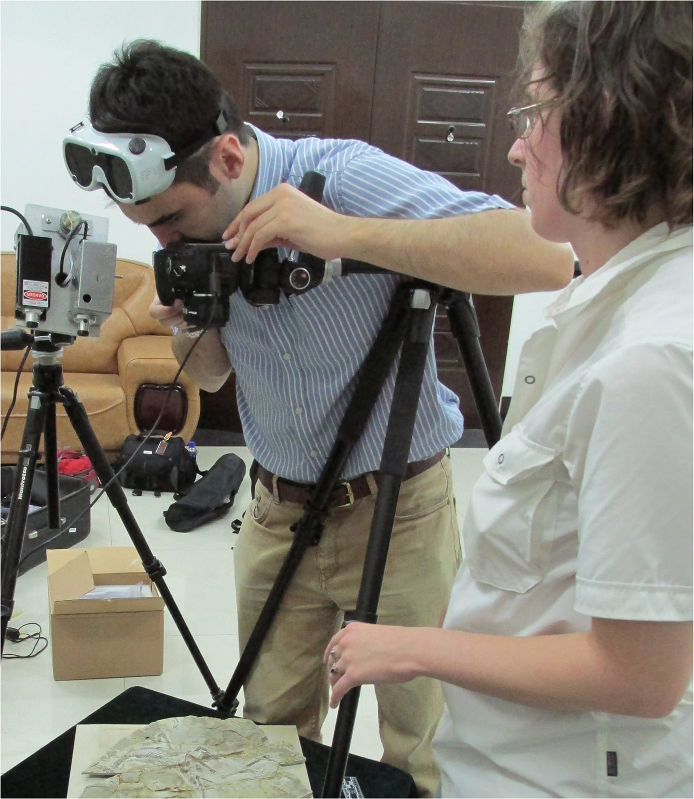
A fossil does not have to glow to be legible. Most are studied first under ordinary light, with the eye tracing bone against stone. Laser-stimulated fluorescence, or LSF, begins when that familiar view stops being enough. In a darkened workspace, a narrow band of intense visible light is swept across the specimen while a camera looks through a filter that blocks the laser itself. Materials that absorbed the incoming energy can emit light at other wavelengths. Differences that were faint—or invisible—under white light become photographable.[1]
The result can feel uncannily complete: the edge of a foot pad, a feather's barbules, a crustacean limb continuing beneath a skim of limestone. Yet the most important discipline arrives before the anatomical excitement. LSF produces a contrast map, not a chemical answer. Brightness and colour can show that two areas respond differently to one combination of laser, filter and exposure. They do not, by themselves, name the molecule or mineral responsible, prove that a halo is original tissue, or turn a soft-tissue outline into a behaviour.[1][2][5]
That boundary is what makes the method useful rather than magical. It tells researchers where the fossil may hold more information, then hands the harder questions to anatomy, taphonomy, chemistry and comparison.
The photograph is built from rejected light
Fluorescence is an energy handoff. A material absorbs incoming photons, some electrons move into excited states, and part of that energy is later emitted as lower-energy light. Fossils are complicated targets because bone, shell, carbon films, surrounding minerals, consolidants and repair adhesives may all contain different luminescent centres. Diagenesis—the chemical history after burial—can alter those centres again. There is no universal “fossil colour” waiting for a laser to switch on.[1][2]
The optical setup turns that complexity into usable contrast. The laser supplies high irradiance at a selected wavelength, often violet, blue or green. A long-pass filter in front of the camera rejects the shorter-wavelength excitation beam while allowing longer-wavelength fluorescence to reach the sensor. For a large slab, the beam can be spread into a line and mechanically swept across the surface during a long exposure. Microscopic work can concentrate it into a smaller spot. Either way, the specimen is usually photographed in darkness so ambient light does not bury the weak emission.[1][2]
In the 2017 study of the feathered paravian Anchiornis, researchers used 405- and 532-nanometre lasers, a blocking filter and repeated sweeps during the camera exposure. The resulting images were adjusted for sharpness, colour balance and saturation.[2] Those processing steps do not invalidate the record, but they do matter. An LSF photograph is not the fossil's “true colour.” It is a deliberately isolated signal whose appearance depends on excitation wavelength, filter cutoff, exposure, camera response and documented post-processing.
A strong workflow therefore keeps comparisons attached. Photograph the same area under white light, ultraviolet illumination and LSF. Include a scale in the specimen plane. Record laser power, wavelength, filter and exposure. Apply any image adjustment consistently across the field. Wear wavelength-appropriate eye protection and control reflections. The method is non-destructive in the sense that it need not cut or sample the fossil; it is not casual illumination.[1][3]
Sometimes the fossil is the dark part
One of LSF's clearest tricks is really a reversal. A carbonized structure may fluoresce weakly or not at all, while the matrix behind it glows strongly. The fossil then appears as a dark silhouette against luminous rock. In the foundational 2015 study, this backlighting made fine barbules visible across Green River Formation feathers where ordinary reflected light showed mainly the larger barbs.[1]
That distinction matters. The laser did not make the feather substance blaze like a neon sign. It made the surrounding material provide a brighter background. Anatomy emerged from negative contrast. Calling every dark edge “soft tissue,” however, would simply move the mistake into a more dramatic colour palette. Cracks, preparation boundaries and non-fluorescent minerals can also interrupt a glowing field. Continuity across known anatomy, repeated occurrence in comparable specimens and agreement with the white-light surface are what make the silhouette persuasive.
The same principle can reveal shallow buried detail. Luke Barlow and colleagues compared LSF with long-wave ultraviolet imaging across fossils from the Upper Jurassic Solnhofen limestones of Bavaria. Decapod crustaceans yielded appendage outlines and segmentation that were incomplete under ordinary or UV light, including portions just beneath the surface.[3] “Beneath” needs its adjective: shallow. LSF can exploit a thin, partly translucent matrix and scattered light, but it does not see through a nodule the way X-ray computed tomography can. The host rock, burial depth and optical properties decide where the beam stops being informative.[1][3]
This is a recurring methodological gain. LSF does not replace a more powerful scanner in miniature. It gives researchers a fast surface and near-surface survey, particularly useful when a slab is too large for a conventional scanner or when fossil and matrix have too little density contrast for a clear X-ray result.[1]
A bright boundary can be a warning
Not every useful fluorescence pattern adds anatomy. Some expose a specimen's later history. Adhesives, fillers and consolidants may respond differently from original matrix. Separate bone fragments may carry unlike mineralization histories. The 2015 paper used a Microraptor skull to show how colour and fluorescence changes across a break could flag a possible composite, although the authors kept an alternative open: different burial conditions might also produce the mismatch.[1]
That is the right evidentiary posture. LSF can identify a boundary worth investigating; it cannot authenticate a fossil on colour alone. Preparation records, microscopy, elemental mapping, mineral analysis and provenance still have to decide whether the contrast marks glue, restoration, separate burial histories or natural variation within one slab.
The method also works as triage. The original experiments showed that high-flux laser illumination could help pick fluorescent microfossils from sediment concentrates.[1] At the other scale, Michael Pittman and colleagues examined more than 1,000 fossils of early theropod flyers and found only 12 specimens with notably preserved toe pads, foot scales and claws. Two famous Archaeopteryx specimens produced no comparable pedal soft-tissue signal.[4]
That ratio is more instructive than a gallery of successes. LSF can search a large collection for rare candidates without claiming that every specimen should respond. A non-fluorescent foot does not prove the living animal lacked pads or scales. It can mean those tissues decayed, their chemical traces were not retained, the matrix masks them, or the chosen excitation-and-filter combination did not separate them. No glow is an analytical result, not an anatomical absence.
Anatomy still requires a second argument
The Anchiornis survey makes the interpretive ladder visible. LSF recovered high-fidelity outlines around the arms, legs and tail, but the head, neck and torso could not be mapped reliably. The authors also stated that they had not chemically determined whether the fluorescent material was organic or mineral.[2] They could describe a spatially coherent body margin without pretending to know the exact substance carrying that margin.
Functional claims required more. Foot-pad grooves had to align with bones and repeat across specimens. Skin textures had to preserve consistent relationships to digits. Comparisons with living birds and other theropods supplied names for structures and bounded possible functions. Even then, an outline of a pad is evidence of anatomy, not a recording of one animal perching, hunting or walking. Behaviour sits another inferential step away.[2][4]
Preservation also chooses the sample before analysis begins. The Solnhofen work found that different fossils, tissues and matrices reacted differently to ultraviolet and laser excitation.[3] A technique that excels in a fine-grained Konservat-Lagerstätte may be unrewarding in a weathered bonebed. If researchers publish only the glowing exceptions, LSF can make the fossil record look more uniformly soft-tissue-rich than the collection survey actually was.
The remedy is not to distrust the image. It is to keep denominators, failures and specimen context beside it: how many fossils were screened, which formations responded, what wavelengths were tried, which regions remained equivocal, and whether the same boundary survives other imaging or chemical tests.
The next instrument asks what made the map
The chemistry gap is now an explicit research target. In 2026, Thomas Kaye and Michael Pittman reported first results from a portable system combining time-gated fluorescence spectroscopy and imaging. Their premise is revealing: ordinary fluorescence imaging can detect parts-per-million-level contrasts in fossils, but the sources of those signals often remain unknown. Time-resolved spectral measurements are intended to add information that a colour photograph cannot supply and to detect certain elements qualitatively without destructive sampling.[5]
This is a bridge, not a universal decoder. The study presents first results from a portable instrument; it does not turn every fluorescent colour into a unique molecule or erase the effects of diagenesis. Its importance is conceptual. Once LSF has located an anomaly, spectroscopy can begin asking what kind of emitter produced it. Spatial discovery and material identification become adjacent measurements instead of one overloaded image.[5]
That sequence preserves the wonder without surrendering the method. A dim fossil enters a dark room. A laser and filter make a boundary visible. Repeated anatomy decides whether the boundary belongs to a body. Chemistry tests what material carries it. Comparative biology asks what the structure could do. Each step narrows a different uncertainty.
The glow is therefore neither decoration nor verdict. It is an invitation to look again—precisely enough to know which question comes next.
Sources
- Thomas G. Kaye et al., “Laser-Stimulated Fluorescence in Paleontology,” PLOS ONE 10 (2015)—foundational setup, fossil–matrix contrast, shallow subsurface imaging, microfossil sorting and molecular-identification limits.
- Xiaoli Wang et al., “Basal paravian functional anatomy illuminated by high-detail body outline,” Nature Communications 8 (2017)—Anchiornis imaging protocol, recovered and unresolved body regions, image processing and the chemistry boundary.
- Luke A. Barlow et al., “Laser-stimulated fluorescence reveals unseen details in fossils from the Upper Jurassic Solnhofen Limestones,” Royal Society Open Science 8 (2021)—controlled LSF-versus-UV comparison and shallow subsurface detail in decapods.
- Michael Pittman et al., “Exceptional preservation and foot structure reveal ecological transitions and lifestyles of early theropod flyers,” Nature Communications 13 (2022)—collection-scale screening, rare pedal soft-tissue preservation and the handoff from imaging to comparative inference.
- Thomas G. Kaye and Michael Pittman, “Elemental analysis of fossils using portable time-gated fluorescence spectroscopy and imaging,” Spectrochimica Acta Part A 363 (online 2026)—first portable-system results and the unresolved-source problem in fossil fluorescence.
- The University of Hong Kong, “New laser-induced fluorescence techniques uncover never-before-seen details in fossils” (2015)—institutional account and source for the documentary setup photograph.