The Herefordshire Lagerstatte is the opposite of a fossil site that wins the reader immediately. It does not offer a giant mounted skeleton, a bedding plane crowded with obvious silhouettes, or a tourist-friendly wall of dramatic bodies. Its classic objects are small marine invertebrates sealed inside nodules from a mid-Silurian volcaniclastic layer, about 430 million years old.[1][3] Many are only millimetres to a few centimetres across.[2] The field report starts there because the site's scientific power is not scale. It is how much anatomy survived inside such unshowy packages.
That makes Herefordshire a useful correction to the way exceptional preservation is often remembered. The Cambrian soft-bodied sites get the public language of explosion and first appearance. Herefordshire asks a quieter question: what did marine invertebrate bodies look like long after the Cambrian, in a Silurian world whose soft-bodied faunas are much rarer in the fossil record?[1] The answer comes through nodules, calcite, volcanic ash, and a deliberately destructive method that turns an invisible fossil into a virtual one.
The first thing to keep straight is the setting. Oxford University Museum of Natural History describes the deposit as a globally important mid-Silurian fossil assemblage, with fossils hosted in nodules in a volcaniclastic layer and preserved with soft parts "in the round."[1] The later Journal of the Geological Society synthesis frames the same point more technically: the site is a rare Silurian example of soft-tissue preservation, yielding a wide range of invertebrates in unusual three-dimensional detail, with fossils preserved as calcitic void infills in early diagenetic carbonate concretions within a bentonite horizon.[3]
That sentence is dense because the preservation is doing several jobs at once. Volcanic material helped build the burial context. Carbonate concretions grew early enough to hold the bodies. Calcitic infill preserved forms that would normally collapse, rot, or vanish. The result is not a normal shell bed with a few lucky soft impressions. It is a small-object archive where sponges, echinoderms, brachiopods, worms, molluscs, arthropods, ostracods, and stranger forms can retain legs, eyes, gill filaments, antennae, spines, and other structures far finer than the ordinary Silurian record usually grants.[2]
The second thing to keep straight is that Herefordshire fossils are hard to read precisely because they are preserved so well. A fossil and its nodule are both mostly calcium carbonate, so the specimen cannot simply be freed with acid without destroying the object being studied.[2] Mechanical preparation is also too crude for many of these tiny, delicate bodies. Even high-resolution CT has historically struggled to distinguish fossil from enclosing rock clearly enough for the standard non-destructive route to work.[2]
That is why the site's signature method feels almost paradoxical. Researchers grind the fossil away in increments, photograph each new surface, and convert the stack of two-dimensional sections into a three-dimensional digital model.[1][2] The Oxford Museum blog describes increments of 20 microns, with each newly exposed surface photographed under a microscope before software turns the image stack into a rotatable virtual fossil.[2] In ordinary museum imagination, preparation reveals an object. At Herefordshire, preparation consumes one physical object so a digital anatomical object can be made from it.
This is not a gimmick. It changes the kind of paleontology the site can do. A flattened fossil can show outline, proportion, and sometimes a soft-tissue stain. A serially reconstructed Herefordshire fossil can expose appendage arrangement, ventral surfaces, mouthparts, limb branching, internal rings, eggs, and soft anatomy that would otherwise remain locked inside the nodule.[2][4][5] The loss is real: the sectioned fossil is gone as a whole specimen. The gain is also real: the resulting model can be rotated, virtually dissected, colored by structure, and compared with living and fossil relatives in a way the original nodule could not allow.[1][2]
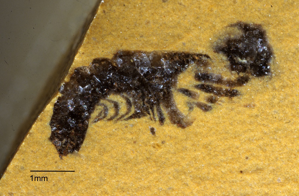
Offacolus makes the method concrete. The fossil in the lead image is not impressive in the way a complete dinosaur is impressive. It is a small chelicerate arthropod from the Wenlock Series of Herefordshire, important because serial grinding and computer reconstruction let researchers render the animal in three dimensions and place it in a phylogenetic argument about early chelicerates.[4] The point is not that the fossil looks like a modern horseshoe crab, scorpion, or spider. The point is that its appendages and body plan can be inspected closely enough to discuss where it sits near that larger branch of arthropod history.[4]
Cascolus ravitis shows the other side of the same archive. Described as a three-dimensionally preserved crustacean with soft parts from the Herefordshire Lagerstatte, it keeps the site from becoming an "Offacolus place" in the reader's mind.[5] Herefordshire matters because it repeatedly turns tiny invertebrates into anatomical case studies. One fossil can sharpen chelicerate questions. Another can sharpen crustacean and mandibulate questions. Others have been used to discuss sea cucumbers, molluscs, ostracods, sea spiders, trilobites, and panarthropod diversity.[2][5]
That breadth is why a field-report mode fits better than a single-species profile. The field lesson is not "look at this animal." It is "look at this preservation-and-method system." Herefordshire sits in a post-Cambrian interval where soft-bodied marine faunas are scarce compared with the famous Cambrian windows, yet the deposit preserves enough soft anatomy to test evolutionary stories that cannot be answered from shells alone.[1][3] In that sense it fills a different gap from Burgess, Chengjiang, or Sirius Passet. It does not dramatize the first burst of animal architecture. It makes a later marine world more bodily.
The caution is just as important. A virtual reconstruction is not raw life restored. It is an interpretation built from hundreds of ground surfaces, each shaped by preservation, grinding precision, imaging, segmentation, and anatomical comparison.[2] The colors in published models are explanatory choices, not original animal colors. A rotatable 3D object can feel more complete than the evidence really is. A good Herefordshire reading therefore keeps the nodule in view even after the model appears. The digital fossil is powerful because the physical process was disciplined, not because software bypassed uncertainty.
That discipline is what makes the site so valuable in 2026. Paleontology increasingly depends on methods that convert difficult material into inspectable data: CT scans, synchrotron imaging, photogrammetry, surface scanning, isotopes, thin sections, and serial grinding. Herefordshire is an especially honest version of that shift because the tradeoff is visible. To recover the animal, researchers may have to destroy the enclosing fossil slice by slice. The archive is not a storehouse of intact treasures waiting to be lifted out. It is a set of nodules that become evidence only when their internal order is patiently converted into another form.[1][2]
The strongest reading of Herefordshire is therefore modest and large at the same time. Modest, because the specimens are small, the surfaces are plain, and every virtual model rests on method. Large, because those tiny nodules preserve a Silurian marine fauna with soft anatomy in three dimensions, at a time when such windows are rare.[1][3] The site turns paleontology away from spectacle and toward procedure: ash buries, carbonate protects, calcite records, grinding reveals, software rebuilds, and the animal finally becomes legible.
Sources
- Oxford University Museum of Natural History, "Soft Bodied Sensations from the Herefordshire Lagerstatte" - institutional event page summarizing the site's age, nodules, volcaniclastic layer, soft parts, and physical-optical tomography.
- Oxford University Museum of Natural History blog, "Revealing Exceptional fossils, one layer at a time" - source for the real Offacolus fossil photograph, nodule context, serial grinding method, and 20-micron image-stack workflow.
- Derek J. Siveter, Derek E. G. Briggs, David J. Siveter, and Mark D. Sutton, "The Herefordshire Lagerstatte: fleshing out Silurian marine life," Journal of the Geological Society 177, no. 1 (2020).
- Mark D. Sutton, Derek E. G. Briggs, David J. Siveter, and Derek J. Siveter, "The arthropod Offacolus kingi (Chelicerata) from the Silurian of Herefordshire, England: computer based morphological reconstructions and phylogenetic affinities," Proceedings of the Royal Society B 269 (2002).
- David J. Siveter, Derek E. G. Briggs, Derek J. Siveter, Mark D. Sutton, and David Legg, "A new crustacean from the Herefordshire (Silurian) Lagerstatte, UK," Proceedings of the Royal Society B 284 (2017).