
A fossil does not photograph itself. Move a lamp a few centimetres and a nearly flat ridge becomes a sharp boundary; raise the light and the same ridge may dissolve into the stone. Change the lens, camera angle, background, or stack of exposures and the object remains physically unchanged while the evidence available to a viewer changes.
That is the productive tension inside Andre Gogol's Royal Tyrrell Museum lecture on fossil photography. Gogol is interested in images that are both anatomically informative and visually arresting. His photographs formed Perspectives, a museum exhibition of detailed views of specimens from the Royal Tyrrell collections.[1][2] The lecture is therefore not a neutral equipment tutorial. It is a demonstration of how photographic choices decide which facts become legible—and which choices a responsible viewer should keep in mind.
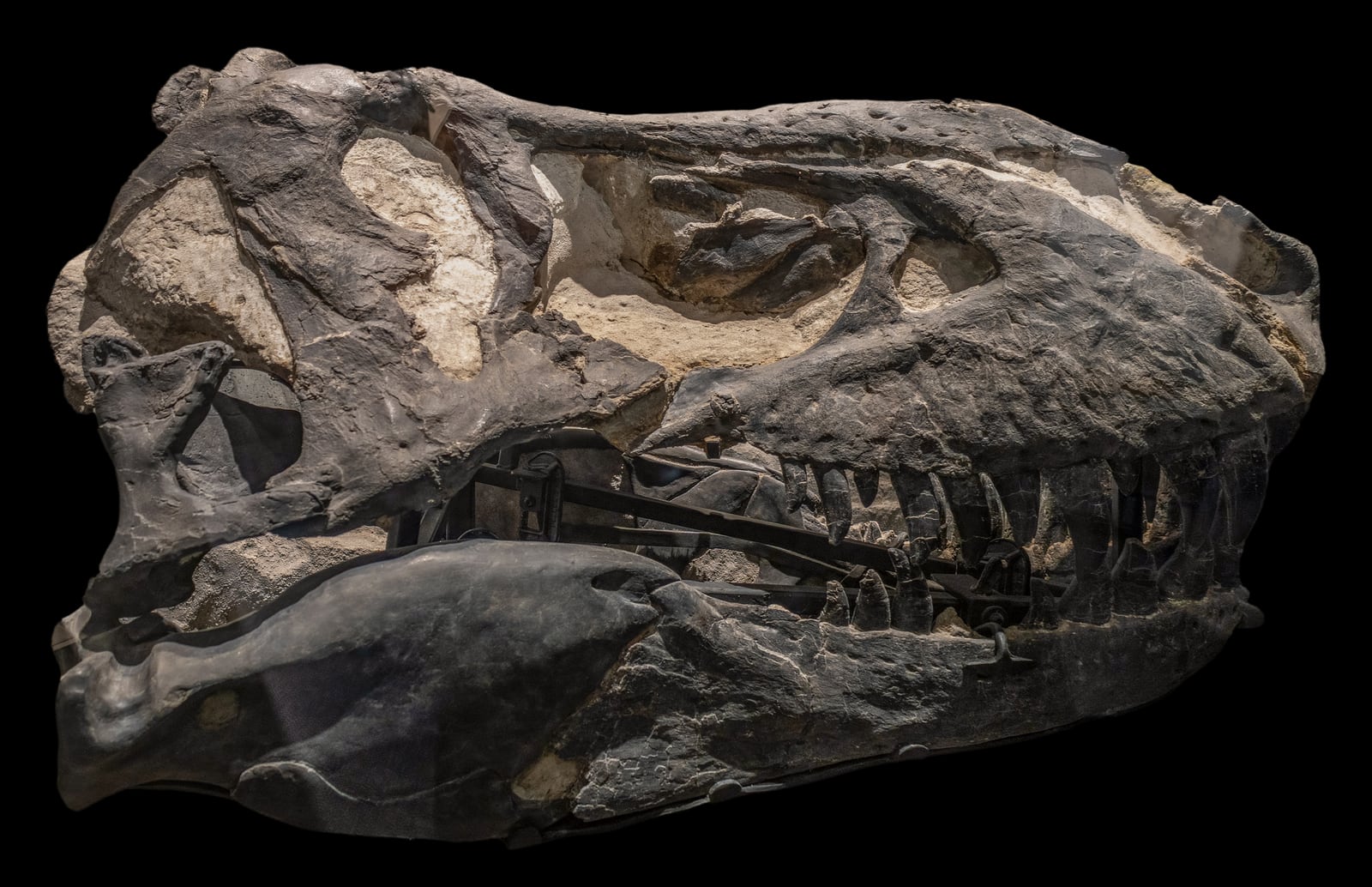
The photograph above supplies a useful object lesson. Black Beauty's long profile is isolated against black, while angled illumination picks out cracks, teeth, and changes in surface texture. It is a compelling encounter with a famous Tyrannosaurus rex skull, but it is not a measurement-ready specimen plate. That difference is central to reading Gogol's work.[7]
The viewing
Light is not decoration; it is a test
At 50:44, Gogol opens the dedicated lighting section by calling light the photographer's “paintbrush.” Watch what happens as he changes its direction and hardness. The useful question is not “Which version looks best?” but “Which kind of structure does this version allow me to test?” Light arriving from near the camera tends to suppress shallow topography. Low, oblique light casts small shadows, so fine relief can become easier to follow. A reflector or a broader source can then soften shadows that have become so dark they hide rather than reveal.[1]
There is no universally truthful lamp position. Hans Kerp and Benjamin Bomfleur's practical study of fossil photography reaches the same conclusion: oblique illumination can make slight relief visible, yet structures expressed mainly by colour rather than height may fade as the angle becomes more extreme.[3] One setup may clarify a suture; another may distinguish a stain from surrounding matrix. The correct response is often a controlled series, not a hunt for one authoritative glamour shot.
This changes how the viewer should read the lecture's dramatic images. Deep black backgrounds and sharply directed light isolate a skull or tooth from its surroundings, giving shape immediate visual force. That can be excellent exhibition photography. For comparison and diagnosis, however, a second image under flatter or differently oriented illumination may carry more information. A photograph is strongest when its purpose is explicit.
Scale belongs to the observation
At 53:29, Gogol describes a detailed specimen image as scientifically accurate, then immediately notes its missing scale bar. The qualification matters. Pay attention to everything else that looks boring: support, alignment, reference scale, and the edge of the frame. Those details determine whether another person can compare the image with another specimen or recover the size and orientation of the object.[1][4]
A scale bar is not a scientific charm. It has to lie in the relevant plane, remain readable, and correspond to the image as reproduced. The camera should not tilt so strongly that the near end of a long fossil is enlarged relative to the far end. Orientation and specimen identity also need to travel with the file. Without them, a highly resolved image may still be weak evidence: it shows texture beautifully but leaves the viewer unsure what, how large, and from which direction.
The lesson is broader than documentation protocol. Framing is an anatomical claim. Cropping out the broken edge of a bone may create a cleaner composition while hiding the boundary between preserved surface and restoration. Turning a specimen to its most imposing angle may conceal the view that would separate two competing identifications. One photograph can invite attention; a defensible record usually needs several views made for comparison.
Focus stacking trades blur for seams
At 39:45, Gogol begins the lecture's most consequential technical explanation: focus stacking. Close-up and microscope photography have shallow depth of field, so when one surface is sharp, a higher or lower surface may blur. A stack records a sequence of focal planes, and software selects sharp regions from those frames to construct one image that appears focused from front to back.[1][3] In microfossil work, the related method called polyfocal photography can reveal structures such as conodont basal cavities and internal white matter more clearly than a single focal plane.[5]
Pause at that transition from raw frames to finished composite. The result is not one exposure with magically improved optics. It is a derived image assembled across space and, usually, time. That is valuable precisely because it solves a real optical limit. It can also create seams around overlapping structures, choose the wrong plane along a translucent edge, or sharpen dust and preparation marks until they compete with anatomy. Kerp and Bomfleur warn that automatic stacking can introduce such artifacts, particularly where software has trouble deciding which region belongs to the object.[3]
The proper conclusion is not that stacked images are suspect. It is that their provenance matters. Retain the source frames. Record the lens, magnification, illumination, spacing between focus levels, and processing method. Inspect the composite beside individual frames before treating a strange ridge as morphology. A repeatable composite is evidence; an unexplained composite is an assertion.
The diagnostic plate and the portrait have different jobs
Later in the lecture, the finished photographs invite a false choice between science and art. A visually forceful fossil image need not be scientifically empty, and a documentary image need not be ugly. What matters is whether the image is asked to do the job it was made to do.
Robert Clark's photographs of the armoured dinosaur Borealopelta at the Royal Tyrrell Museum offer a useful comparison. He used deliberate lighting to reveal the fossil's extraordinary surface while also producing images capable of carrying the specimen to a public that could not examine it outside glass.[6] That public encounter is a legitimate function. It rewards isolation, scale, and drama. A taxonomic figure may instead prioritize even exposure, multiple orientations, labels, and a visible scale. Neither format cancels the other; trouble begins when the portrait is mistaken for a complete technical record.
The Black Beauty photograph makes the distinction tangible. Its raking light and black ground turn the famous skull into a continuous, forceful silhouette. There is no scale or specimen number inside the frame, and the oblique perspective privileges the profile over exact proportion.[7] Those choices are not defects in a public-facing portrait. They would become defects only if the portrait were offered as the sole technical record. The image's job and its evidentiary boundary belong together.
The best way to leave the lecture is to stop treating photography as the last cosmetic step after palaeontological work. Illumination can test relief. Scale and orientation make comparison possible. Focus stacking can recover visibility across depth while introducing a new class of artifacts. Framing assigns importance. A strong fossil photograph does not pretend to replace the specimen; it makes a particular observation transportable and leaves enough of its method visible for someone else to question it.
Sources
- Royal Tyrrell Museum of Palaeontology, “Exploring Methods in Fossil Photography,” lecture presented by Andre Gogol on February 26, 2025, YouTube.
- Royal Tyrrell Museum of Palaeontology, Annual Report 2024, noting the opening of Perspectives and its detailed photographs of specimens from the museum's collections.
- Hans Kerp and Benjamin Bomfleur, “Photography of plant fossils—New techniques, old tricks,” Review of Palaeobotany and Palynology 166 (2011), pp. 117–151.
- Sam Noble Oklahoma Museum of Natural History, “Photographing Macrofossils Procedures,” museum imaging protocol for scale, specimen identifiers, camera alignment, and lighting.
- James F. Miller, Benjamin F. Dattilo, Raymond L. Ethington, and Rebecca L. Freeman, “Polyfocal photography of conodonts and other microfossils using petrographic microscopes,” Revue de Micropaléontologie 58 (2015), institutional record.
- National Geographic, “This Dinosaur Is the ‘Most Impressive Fossil’ We've Ever Seen,” photographic account of Borealopelta at the Royal Tyrrell Museum.
- Chris Woodrich, “Black Beauty, Royal Tyrrell Museum of Palaeontology, Drumheller, Alberta,” 2025 photograph, Wikimedia Commons, CC BY-SA 4.0.