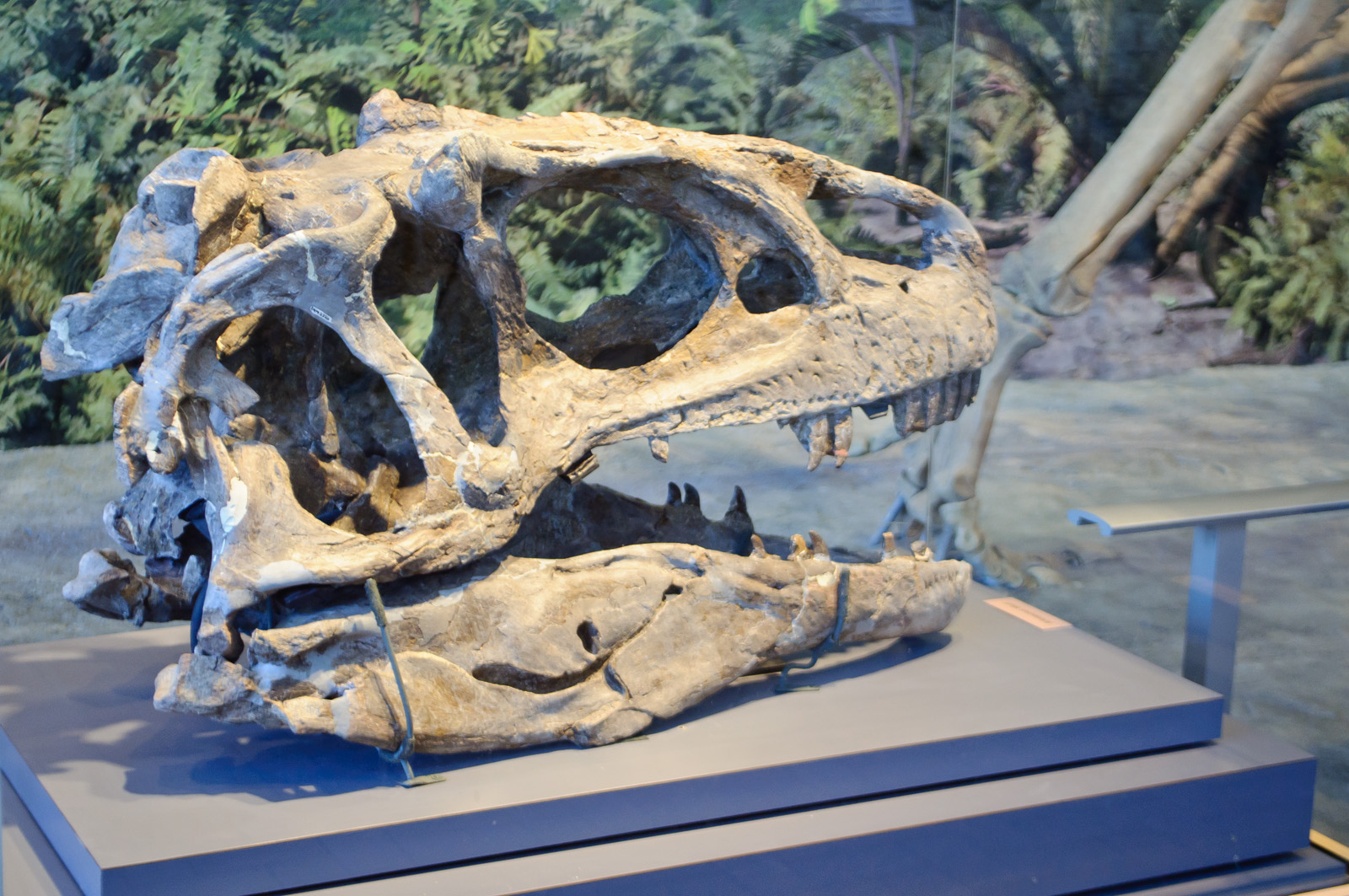
A fossil skull arrives as the aftermath of several possible transformations. Flesh vanished. Sediment may have filled openings; mineralization may have altered tissue; individual bones may have cracked, shifted, flattened, or disappeared. Museum preparation then made the surviving anatomy legible again.[1]
Then a second transformation can begin. A researcher scans the skull, repairs it in three dimensions, divides the restored volume into a mesh, gives that mesh mechanical properties, fixes it at imagined joints and bite points, and pulls on it with reconstructed muscles. Software calculates how the digital structure deforms. The result often arrives as a spectacular contour map: cool blues across lightly stressed regions, hot yellows and reds where stress concentrates.
That rainbow skull is not an X-ray of an extinct animal biting. It is the answer to a conditional question: if this reconstruction had these material properties, and if these forces acted in these directions while these points were constrained, how would the structure respond? Finite-element analysis, or FEA, is powerful precisely because those conditions can be changed one at a time. Its rigor lives in the conditions, not in the colors.[2][3]
Image context: the cover is a real photograph of a fossil Allosaurus skull on display, not a stress visualization or life reconstruction. The National Park Service identifies the remarkably preserved skull as a real fossil from Carnegie Quarry.[10] It supplies the preserved anatomical object. The analytical chain described below begins only after that object is digitized and interpreted.[1][3]
The model begins before the mesh
FEA originated as an engineering way to estimate the behavior of complicated structures under load. In paleontology, however, the first engineering problem is often not force. It is shape.
A scan records the specimen as preserved, including damage. That may be exactly what a taxonomic description needs, but it is not necessarily the geometry of a living skull. One side may be crushed inward. A snout may have twisted during burial. Sutures can be obscured by matrix, and an entire cheek bar may be absent. Before testing mechanics, researchers may digitally separate bones, close cracks, rearticulate displaced elements, mirror a better-preserved side, or borrow missing geometry from another specimen or a close relative.[1]
None of those moves is automatically illegitimate. A mirrored bone can be a more defensible estimate than leaving a taphonomic hole in a structure meant to carry force. But every repair adds a proposition. Mirroring assumes enough bilateral symmetry. Repositioning assumes the joint has been identified correctly. Borrowing from a relative assumes that the substituted shape is mechanically appropriate. Digital restoration can recover information that stone hides while also making interpretation look seamless.[1]
The cleanest studies preserve that seam. They document which regions are original, restored, mirrored, or borrowed; retain alternative reconstructions when more than one is plausible; and ask whether the mechanical conclusion survives the difference. A model with two defensible skull shapes is not a failure to decide. It is an experiment with anatomy as a variable.[1][3][5]
From skull to solvable pieces
Once the surface is restored, the continuous skull is subdivided into many simple, connected elements. The corners of those elements are nodes. Taken together, they approximate a shape too complex to solve as one object. Increasing element count can improve the representation of thin bars, curved sutures, and internal cavities, but a denser mesh is not automatically a truer model. Researchers use convergence tests to check whether further refinement materially changes the answer.[2][3]
The mesh is only geometry until it receives a mechanical world. Each part needs elastic properties describing how much it resists deformation. The model needs loads: perhaps forces estimated for jaw-closing muscles, an impact, or a bite. It also needs constraints that stop the skull from drifting freely through virtual space—commonly at the jaw joints and the tooth contacting food. The solver then estimates displacement, strain, and stress throughout the structure. Strain describes relative deformation; stress describes internal force per unit area within the material.[2][3]
For a bridge, an engineer can test the steel and know where a support touches the ground. An extinct skull does not offer those luxuries. The mechanical properties of living bone cannot be measured directly in fossilized tissue, so model values come from living analogues or justified approximations. Jaw muscles are absent. Their size and direction must be reconstructed from attachment areas, close living relatives, and assumptions about muscle architecture.[2][3] A tooth mark may establish that a bite happened, but it does not preserve every muscle’s activation at that instant.
This is why an FE model is not simply “the fossil in a computer.” It is a fossil-derived geometry inside an explicitly built mechanical scenario.
Material can preserve the pattern while moving the numbers
Validation exposes the difference between a useful pattern and a trustworthy magnitude. In a 2011 study, researchers loaded a modern pig skull, measured strain with gauges, and compared those measurements with specimen-specific FE models subjected to equivalent conditions. The models generally recovered the orientation and broad distribution of strain, but the first homogeneous model was roughly an order of magnitude too stiff. Adding a more compliant interior improved the magnitudes without greatly changing the contour pattern.[4]
That is an encouraging result with a sharp boundary. A simplified model may locate the parts of a skull that work relatively harder even when its absolute strain values are wrong. For extinct animals, whose living bone properties cannot be measured, the distinction becomes decisive. A model may support “shape A distributes this standardized load more evenly than shape B” more securely than “this skull would break at exactly this force.” Reviews of paleontological FEA therefore emphasize relative comparisons and sensitivity tests, especially when absolute failure or bite-force claims depend on poorly constrained tissue properties.[3][4]
Greater complexity is not a universal cure. Assigning a dozen material regions is useful only if the divisions and property values have evidence behind them. A beautifully heterogeneous model can encode more unsupported precision than a simple one. The relevant question is not how detailed the model looks, but whether added detail changes the inference and whether that detail can be justified.
Behavior enters through the loading case
Two identical meshes can tell different stories when loaded differently. A front-tooth bite, a rear-tooth bite, a pull, and a sideways shake route forces through different parts of a jaw. Muscle force applied across a broad attachment is not mechanically identical to the same total force concentrated at one node. Locking a joint in too many directions can create artificial stiffness; leaving it too free can create motion the living joint would not permit.[2][3][5]
A seven-species crocodilian study varied material properties, scaling choices, tooth positions, and loading cases. Its comparative rankings were less sensitive to some reasonable changes in material properties than to functional choices such as which tooth engaged and what kind of load was simulated. Crucially, applying the same assumptions to every species did not guarantee that the result generalized beyond those assumptions: different skull shapes interacted with the load cases in different ways.[5]
That lesson is especially important when behavior is the conclusion. If one modeled bite makes a fossil taxon look unusually resistant, the safe statement concerns that bite configuration. Claims about prey capture, head striking, shell crushing, or feeding ecology require additional anatomical, wear, trace-fossil, or comparative evidence. FEA can make a scenario less plausible under the tested assumptions—especially when poor performance persists across sensitivity tests—or show that one shape performs differently from another.[2][3][5] Mechanical possibility alone does not prove that an animal habitually used the behavior.
Comparison is stronger when size is handled openly
Large skulls and small skulls can be subjected to the same force, but that equal-load experiment does not isolate shape by itself. Size affects cross-sectional area, lever arms, muscle capacity, and the amount of material available to carry load. A larger structure may withstand more absolute force even if its shape is less mechanically efficient.
One influential framework showed how scaling models to an equivalent force-to-surface-area relationship can remove the direct effect of size from stress comparisons, allowing shape to be tested more cleanly. The same work proposed total strain energy—the work expended deforming a structure—as a comparative measure of mechanical efficiency.[6] Neither operation reveals a single “best” skull. It states which performance question is being isolated.
This creates two legitimate but different experiments. Actual-size models with biologically estimated loads and constraints ask how the animals’ skulls may have performed with their biological scale retained. Size-standardized models ask what their shapes would do if scale were held comparable. Confusing those experiments can turn an efficient shape into a claim about absolute strength, or a giant’s high absolute capacity into a claim about superior design.
Eighteen theropods, two questions about gigantism
A 2025 study illustrates what FEA can do when the comparison is made explicit. Researchers assembled three-dimensional cranial and mandibular models for 18 carnivorous theropods and analyzed feeding loads both at actual size and under size-corrected conditions. Under those modeled cases, non-tyrannosauroid skull stress generally did not increase with size, whereas tyrannosaurids experienced greater cranial stress associated with larger estimated jaw-muscle volumes and modeled bite forces. The authors interpreted those patterns as different mechanical routes to giant predation.[8]
The interesting result is not that software replayed an encounter between Tyrannosaurus and prey. It did not. The study compared virtual structures under declared loading and scaling regimes. Some source geometries came from CT scans, others from surface scans, casts, mounted specimens, or restored models; the methods disclose those differences.[8] The conclusion is therefore strongest at its stated level: within the constructed comparative experiment, theropod lineages did not scale skull performance in one uniform way.
That is a substantial comparative biomechanical result with evolutionary implications. It becomes more informative, not less, when the conditional grammar stays visible.
Even the rainbow is an analytical choice
The final contour plot can introduce one more layer of persuasion. Red seems dangerous; blue seems safe. Yet the colors are labels assigned to numerical intervals, not heat, pain, cracks, or preserved tissue. Similar hues across two figures are comparable only when the plotted metric and scale match.[7]
A 2021 study tested color maps used in paleontological FEA and found that the familiar rainbow scheme represents numerical change unevenly. Abrupt perceptual transitions can make modest differences look like boundaries, bright yellow draws attention despite sitting inside the scale, and red–green deficiencies can make the map inaccessible. Alternative sequential or diverging palettes often represented particular stress data more faithfully, although no single palette was best for every output.[7]
The study did not show that FEA results are arbitrary. The calculated values do not change when their display palette changes. It showed that legibility is part of scientific interpretation. A serious figure needs a stated metric, units or normalization, a shared scale for fair comparison, and ideally numerical summaries alongside the surface colors.
Read the assumptions before the skull
A well-calibrated FEA paper should let a reader reconstruct the experiment in words. Which specimen supplied the geometry? What was repaired, mirrored, warped, or borrowed? Were internal cavities and different tissues represented? How were muscle size and direction estimated? Where was the model constrained, which tooth or surface was loaded, and why? Were differently sized animals compared at actual scale, standardized scale, or both?
Then come the two confidence tests. Sensitivity analysis reruns the model with plausible alternative inputs and asks whether the conclusion survives. Validation compares a specimen-specific model of an extant analogue with physical strain measured under matched experimental loads, testing what the workflow reproduces and where it fails.[3][4][5] Neither can recover a missing dinosaur muscle. Together they can show whether the inference depends on one fragile guess.
Finite-element analysis gives paleontology something fossils alone cannot: a repeatable counterfactual laboratory. A jaw can bite at several positions. A crushed skull can be tested before and after restoration. A crest can be removed without harming the specimen. A small skull can be rescaled to separate geometry from bulk.
The method does not make an extinct animal confess how it lived. It makes assumptions executable. The rainbow skull earns confidence when changing those assumptions does not erase the pattern—and when the conclusion stays no larger than the experiment that produced it.
Sources
- Stephan Lautenschlager, “Reconstructing the past: methods and techniques for the digital restoration of fossils,” Royal Society Open Science 3 (2016) — University of Bristol record for the open review of scanning, retrodeformation, mirroring, reassembly, and the interpretive boundaries of virtual restoration.
- Emily J. Rayfield, “Finite Element Analysis and Understanding the Biomechanics and Evolution of Living and Fossil Organisms,” Annual Review of Earth and Planetary Sciences 35 (2007) — University of Bristol record for the foundational review of FEA, stress, strain, validation, and form–function hypothesis testing.
- Jen A. Bright, “A Review of Paleontological Finite Element Models and their Validity,” Journal of Paleontology 88 (2014) — institutional record and abstract covering input sensitivity, validation, material uncertainty, and the strength of relative over absolute comparisons.
- Jen A. Bright and Emily J. Rayfield, “Sensitivity and Ex Vivo Validation of Finite Element Models of the Domestic Pig Cranium,” Journal of Anatomy 219 (2011) — institutional record and abstract for the physical strain-gauge comparison and material-property tests.
- Christopher W. Walmsley and colleagues, “Beware the black box: investigating the sensitivity of FEA simulations to modelling factors in comparative biomechanics,” PeerJ 1 (2013) — open comparative study varying materials, scaling, tooth position, and crocodilian loading cases.
- Elizabeth R. Dumont, Ian R. Grosse, and Graham J. Slater, “Requirements for comparing the performance of finite element models of biological structures,” Journal of Theoretical Biology 256 (2009) — framework for separating size and shape and comparing strain energy.
- Stephan Lautenschlager, “True colours or red herrings?: colour maps for finite-element analysis in palaeontological studies to enhance interpretation and accessibility,” Royal Society Open Science 8 (2021) — open test of rainbow and alternative contour palettes.
- Andre J. Rowe and Emily J. Rayfield, “Carnivorous dinosaur lineages adopt different skull performances at gigantic size,” Current Biology 35 (2025) — open 18-taxon comparison using actual-size and size-corrected three-dimensional FE models.
- Wikimedia Commons, “Allosaurus skull, Dinosaur National Monument (7063605345)” — source page, authorship, license, and provenance record for the fossil-skull photograph used as the article image.
- U.S. National Park Service, “Allosaurus fragilis” — official specimen page confirming that the remarkably preserved Carnegie Quarry skull is a real fossil displayed in Dinosaur National Monument’s Quarry Exhibit Hall.