A fossil tooth can leave an excavation with two kinds of history attached. Its worn crown records a working mouth. Inside the enamel, changes too small to see record exposure to radiation. Reading that second history begins before the tooth reaches a laboratory: someone must document where it lay and collect the sediment touching it.[1][2]
Electron spin resonance dating, usually shortened to ESR, makes those unglamorous field records part of the clock. The method can investigate Pleistocene mammals beyond the useful reach of radiocarbon. Its promise is direct access to the fossil's history; its difficulty is reconstructing the conditions under which that history accumulated.[3]
What the instrument reads
Tooth enamel contains densely packed crystals of carbonated hydroxyapatite. Natural ionizing radiation produces changes in their electronic structure, leaving unpaired electrons associated with defects. ESR spectroscopy detects these radiation-sensitive centers. With suitable preservation, their accumulated signal carries information about the energy absorbed over burial time.[1][3]
The first result is a dose, measured in grays. To obtain an age, researchers also need a dose rate: how much energy the enamel absorbed per year. If that rate remained constant, the arithmetic would be accumulated dose divided by annual dose. A long exposure to weak radiation can therefore resemble a shorter exposure to stronger radiation. The signal alone cannot distinguish them.[1]
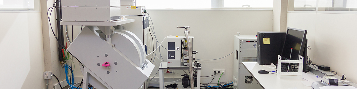
Laboratory irradiation helps establish the accumulated dose. Researchers add known doses and measure how the enamel's signal responds, fitting a response curve from which the earlier exposure can be estimated. CENIEH's laboratory accordingly has separate facilities for irradiation, ESR measurement and environmental radioactivity. The instrument beside the sample supplies only part of the dating evidence.[1][3]
The ground belongs in the calculation
In their 2017 sampling guide, Maïlys Richard and colleagues ask excavators to preserve a tooth's exact position and its surrounding sediment. Nearby radioactive elements contribute to the dose; sediment water content and the thickness of overlying deposits also matter. Their practical recommendation includes collecting at least 150 grams of sediment with the tooth.[2]
That small bag has a different purpose from a museum label. A locality name might identify a valley or cave while leaving the radiation environment poorly constrained. Measurements near the original position help recover that missing information. In uneven deposits, adjacent layers or rock fragments may need attention too.[2]
A tooth moved from one deposit into another is especially troublesome. Its present surroundings cannot automatically stand in for its whole burial history. This is why the guide favors specimens still in their original depositional position: an intact crown may have a complicated geological itinerary.[2]
Uranium arrives after the animal
The tooth itself also changes the radiation budget. Dental tissues can take up uranium during burial, so the amount present today does not reveal how much was present throughout the past. Uranium acquired early has had longer to contribute radiation than uranium acquired late. Combining ESR measurements with uranium-series analyses helps constrain that uptake history.[3]
A Sardinian example makes the consequence tangible. In 2006, Oswaldo Baffa and colleagues reported ESR work on teeth of the extinct bovid Nesogoral melonii from Orosei, in the Nuoro area. Their estimated accumulated dose was 211 ± 34 grays. Assuming early uranium uptake produced an age of 195,000 ± 30,000 years; assuming uptake increased linearly through time produced 247,000 ± 40,000 years.[4]
Both estimates place the material in the Middle Pleistocene. They nevertheless differ by tens of thousands of years because the assumed radiation history differs. These are alternative calculations from the same study, with overlapping uncertainties. Averaging their central values would hide the question the researchers were trying to resolve: how did uranium enter the dental tissues?[4]
A well-shaped tooth can still be altered
Preserving a recognizable molar is not the same as preserving an uncomplicated dating sample. In 2024, Michael Toffolo and Maïlys Richard published an infrared reference library built from modern African ungulate enamel. It provides comparisons for detecting changes in the crystal structure of fossil teeth before committing them to ESR and uranium-series dating.[5]
The comparisons need biological care. Different species begin with different degrees of crystal order, so one modern tooth cannot supply the baseline for every fossil. The library covers fifteen species. Against appropriate references, increased crystal order can flag burial alteration associated with uranium uptake; uranium loss is particularly difficult because it removes information needed to reconstruct the dose history.[5]
This screening does not supply a replacement age. It helps select specimens whose mineral history offers a better chance of a reliable one. The visually handsome tooth and the most useful tooth for dating need not be the same specimen.[5]
The clock can change in the museum
Excavation adds another chapter. Micro-CT scanning uses X-rays to reveal internal anatomy, but those X-rays can also add a radiation dose. CENIEH's direct-dating research explicitly includes measuring this contribution because it can affect subsequent ESR results.[6]
An operation can leave a tooth's shape intact while altering the property another researcher intends to measure. That makes the specimen's analytical history relevant alongside its field history: previous scans belong in the conversation about dating, even when the fossil looks untouched.[6]
Follow the process back from the spectrometer and the apparent simplicity of a tooth date opens into a chain of observations. There is the enamel signal, the sediment, the uranium history, the preservation assessment and the record of laboratory treatment. Each answers a different part of the age question. The small bag of earth collected beside the fossil remains part of the evidence long after the tooth has been cleaned.
Sources
- CENIEH, “Electron Spin Resonance Dating” — laboratory methods, equipment and the photograph reproduced here.
- Maïlys Richard and colleagues, “Electron spin resonance dating of fossil teeth: some basic guidelines to ensure optimal sampling conditions,” Quaternaire 28(2), 155–159 (2017) — institutional record with full text.
- Mathieu Duval, “Dating fossil teeth by electron paramagnetic resonance: how is that possible?,” Spectroscopy Europe 26(1), 6–13 (2014) — dose reconstruction and uranium uptake.
- Oswaldo Baffa and colleagues, “ESR dating of an ancient goat tooth from Nuoro, Sardinia, Italy,” Radiation Protection Dosimetry 119, 446–449 (2006) — abstract and figures reproduced on PDF page 19 of University of Ferrara teaching material.
- Michael B. Toffolo and Maïlys Richard, “Infrared spectral library of tooth enamel from African ungulates for accurate electron spin resonance dating,” Scientific Data 11, 890 (2024) — preservation screening and species-specific reference material.
- CENIEH, “Direct dating of fossils” — uranium screening and research into the effect of micro-CT irradiation on ESR dating.