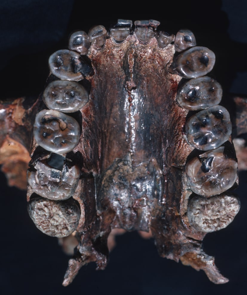
The upper jaw of Paranthropus boisei looks like a machine built around its molars. The cheek teeth are enormous and flat, their enamel is exceptionally thick, and the skull opened room for powerful chewing muscles. After OH 5 was found at Olduvai Gorge in 1959, “Nutcracker Man” became a nickname and, almost inevitably, a menu: nuts, seeds, roots, anything hard enough to justify the apparatus.[2]
But anatomy does not preserve a shopping list. It records an inherited structure that could survive certain loads over a lifetime. On the chewing surface, another archive is being written and erased at a far smaller scale. Food, particles carried with food, opposing teeth and jaw movement leave pits, grooves and directional textures in enamel. Dental microwear texture analysis, or DMTA, maps that relief in three dimensions and compares it with surfaces made by known diets.[1]
The distinction is the method's power and its boundary. A fossil tooth can retain evidence of recent meals; it cannot, by microwear alone, disclose a species' lifelong menu. The surprising case of P. boisei shows why a narrow clock can correct a broad story without replacing it.
A cast carries the surface out of the museum
DMTA begins before a microscope is switched on. A researcher has to choose the correct tooth, the correct wear facet and, most importantly, a surface that still carries marks made during life. A polished patch from sediment transport, an etched patch from chemical alteration or a layer of consolidant may look beautifully measurable while answering the wrong question.
The 2008 P. boisei study made dental impressions of original molars in museum collections, then poured high-fidelity epoxy replicas. This let the researchers examine the same topography without repeatedly handling rare fossils or trying to place an irreplaceable jaw beneath the instrument. They screened the replicas with light microscopy and a confocal profiler before accepting a standard crushing-and-grinding area known as facet 9.[2]
That screening was severe. Of 53 numbered individuals examined, only seven preserved unobscured, clearly pre-death microwear suitable for the analysis. Those seven came from Kenya, Tanzania and Ethiopia and spanned much of the species' known range, from roughly 2.27 million to 1.4 million years ago. OH 5 was among them.[2] Seven is a small sample, but 7 of 53 is also an honest denominator. Rejecting a seductive surface is part of the measurement, not lost productivity.
The sampling geometry must stay equally disciplined. Different parts of a molar do different work as teeth meet and slide. A pit field on a crushing facet cannot be compared casually with scratches on a shearing edge. Studies therefore match tooth position and facet where possible, scan multiple adjacent fields and carry the median or distribution forward. The goal is not the most dramatic patch. It is a repeatable patch whose biological role and preservation history are known.[1][2]
The scanner does not simply count scratches
Earlier microwear studies often used two-dimensional scanning-electron images and asked an observer to identify and count discrete pits and scratches. That produced valuable results, but apparent feature boundaries could shift with magnification, lighting, specimen orientation and the person doing the counting. DMTA replaced much of that judgement with a white-light confocal scan: a point cloud describing the enamel surface's height across a microscopic field.[1]
Scale-sensitive fractal analysis then asks how the surface behaves rather than how many marks a person can name. Complexity describes how roughness changes as it is measured at different scales; a surface cut by pits and scratches of many sizes tends to be more complex. Anisotropy describes how strongly texture shares an orientation; long, similarly aligned striations produce a more directional surface. Other variables can capture heterogeneity, depth or volume, but no single parameter translates directly into a named food.[1]
Meaning comes from comparison. Living primates that process hard, brittle items can provide one reference distribution; animals that repeatedly shear tough leaves or stems can provide another. Fossils scanned with a compatible protocol are located against those empirical patterns. High complexity is therefore not a microscopic photograph of “a nut,” and high anisotropy is not a species label for “grass.” Each is a surface association produced by contacts, movements and materials that must be interpreted through a comparative sample.[1][3]
Microwear is also rapidly overwritten. New contacts cross, soften or remove older features, creating the so-called last-supper effect. In the P. boisei analysis, the authors treated the texture as evidence for foods consumed in the days before death.[2] That makes DMTA unusually intimate, but it also makes a seasonal fallback food easy to miss. A population sample can reveal variation among individuals; one perfect tooth cannot stand for every season of a species lasting hundreds of thousands of years.
The Nutcracker test produced light wear
If the familiar reconstruction were right in its simplest form—if P. boisei routinely crushed hard objects—its molars should repeatedly resemble hard-object feeders in the comparative sample. They did not. All seven accepted specimens had light microwear, generally dominated by fine striations. None displayed the large, deep pits expected of a habitual hard-object specialist, and their complexity values were uniformly low. Nor did they show the extreme directional texture used to characterize habitual processing of very tough foods.[2]
This result removes one claim and leaves several possibilities alive. It argues against those seven individuals having eaten exceptionally hard or tough foods shortly before death. It does not show that no P. boisei ever cracked a hard item. Rare fallback foods might have selected for a robust chewing system yet vanish from a short microwear window. Alternatively, broad molars and powerful muscles could have processed large quantities of lower-quality vegetation without regularly producing a hard-object texture. A small fossil sample cannot choose cleanly between every version of those hypotheses.[2]
Carbon isotopes add a different constraint. Enamel from P. boisei carries a strongly C4-dominated signal, pointing to resources ultimately derived from tropical grasses or sedges rather than the hard-item diet implied by the nickname.[6] Yet isotopes identify a photosynthetic pathway, not texture, plant part or cooking instructions. A sedge stem, underground storage organ and seed can share a broad carbon source while presenting very different mechanical challenges. Microwear and isotopes agree that the original Nutcracker caricature is inadequate; neither turns the replacement into a single food.
This is not a contradiction between good methods. Skull form estimates capacity and long-term selective history. Enamel chemistry integrates resources consumed while a tooth was forming. Microwear samples recent physical contacts on a functioning crown. The clocks differ, so their disagreement is often the discovery.
The mark has more than one possible maker
Even a pristine pre-death scratch is not a direct cast of food tissue. Enamel sits inside a contact system. The intrinsic material properties of a food matter, but so do quartz grit stuck to it, microscopic plant silica, tooth-on-tooth contact, particle shape, force and the path taken by the jaw.
Nanowear experiments sharpened this problem by sliding individual particles across enamel. Quartz dust could fracture and remove enamel under suitable geometry, while softer phytoliths and enamel fragments deformed and produced grooves without the same tissue loss.[3] Controlled feeding experiments then complicated any attempt to declare dust the whole signal: sheep eating browse or grass still developed distinguishable textures even when dust was added, suggesting that environmental particles did not simply erase the dietary contrast under those conditions.[7]
Those findings set a productive boundary. DMTA can discriminate feeding regimes without making every groove chemically unique. A “hard-object” texture may reflect brittle food, adherent grit or both; a directional texture may combine material properties with repeated jaw motion. Researchers gain confidence when experimental baselines, living analogues and independent proxies converge. They should be cautious when a fossil point cloud is asked to name an ingredient by itself.
Burial can write a meal that never happened
Fossilization adds a second contact history. A loose tooth can roll through sediment. Windblown grains can abrade an exposed crown. Acidic conditions can etch enamel, and preparation can add polish or residue. Experiments on modern teeth show that post-death transport and chemical alteration can replace or overprint diet-related textures; the effect varies with particle size and process, and experimental damage is comparable to, though not identical with, natural taphonomic alteration.[4]
This returns us to the 46 rejected P. boisei individuals. The researchers did not average questionable surfaces into a larger, more impressive dataset. They looked for unambiguous pre-death texture on the same functional facet and excluded the rest.[2] A robust analysis records that attrition: which teeth were available, which surface was scanned, why specimens failed, how molds were made and whether the accepted marks cross cracks or preparation boundaries.
Taphonomy cannot always be solved by software. A beautifully quantified post-death surface remains post-death. Context, low-magnification inspection and comparison across a crown have to come before fractal parameters. The most important reading of a fossil tooth may be that it has become unreadable.
Put the clocks beside one another
Dental evidence is strongest as a set of overlapping exposures. Unworn crown shape and jaw architecture describe what an animal was built to process. Mesowear—the macroscopic rounding and relief of molar cusps—accumulates over a longer interval than rapidly changing microwear and can characterize habitual abrasion or attrition in suitable herbivores.[5] DMTA catches the short surface record. Isotopes sample dietary sources during tissue formation. Each proxy has a different unit, timescale and failure mode.
For P. boisei, that stack transforms a mascot into an ecological question. Its skull still mattered; the microwear result did not shrink those molars or muscles. Instead, it forced their meaning away from “this is what the animal always ate” toward “this is what the animal was capable of processing.” The isotope signal then shifted attention toward C4 resources, while the light wear kept food texture open.[2][6]
That is what a good method deep dive should leave intact: not a new nickname, but an inference chain. First establish that the surface belongs to life rather than burial. Measure a homologous facet in three dimensions. Compare distributions, not just spectacular marks. Keep individuals visible inside a species average. Then set the short microwear clock beside anatomy, chemistry, larger-scale wear and environmental context.
A fossil tooth remembers contact. Paleontology earns the menu only by assembling the other clocks around it.
Sources
- Robert S. Scott et al., “Dental microwear texture analysis: technical considerations,” Journal of Human Evolution 51 (2006)—the confocal, three-dimensional and scale-sensitive fractal framework for complexity, anisotropy and repeatable comparison.
- Peter S. Ungar, Frederick E. Grine and Mark F. Teaford, “Dental Microwear and Diet of the Plio-Pleistocene Hominin Paranthropus boisei,” PLOS ONE 3 (2008)—specimen screening, facet protocol, seven-individual result, interpretive limits and source of the OH 5 photograph.
- Peter W. Lucas et al., “Mechanisms and causes of wear in tooth enamel: implications for hominin diets,” Journal of the Royal Society Interface 10 (2013)—nanowear experiments on quartz, phytoliths and enamel and the mechanics linking particles to marks.
- Katrin Weber et al., “Post-mortem enamel surface texture alteration during taphonomic processes—do experimental approaches reflect natural phenomena?” PeerJ 10 (2022)—experimental transport, abrasion and acid alteration of diet-related enamel textures.
- Nicole L. Ackermans, “The history of mesowear: a review,” PeerJ 8 (2020)—how macroscopic cusp relief and shape provide a longer-window wear proxy distinct from microwear.
- Thure E. Cerling et al., “Diet of Paranthropus boisei in the early Pleistocene of East Africa,” Proceedings of the National Academy of Sciences 108 (2011)—carbon-isotope evidence for a strongly C4-based resource signal and its relationship to microwear and morphology.
- Gildas Merceron et al., “Untangling the environmental from the dietary: dust does not matter,” Proceedings of the Royal Society B 283 (2016)—controlled sheep feeding trials testing whether added dust overwhelms diet-related microwear texture.