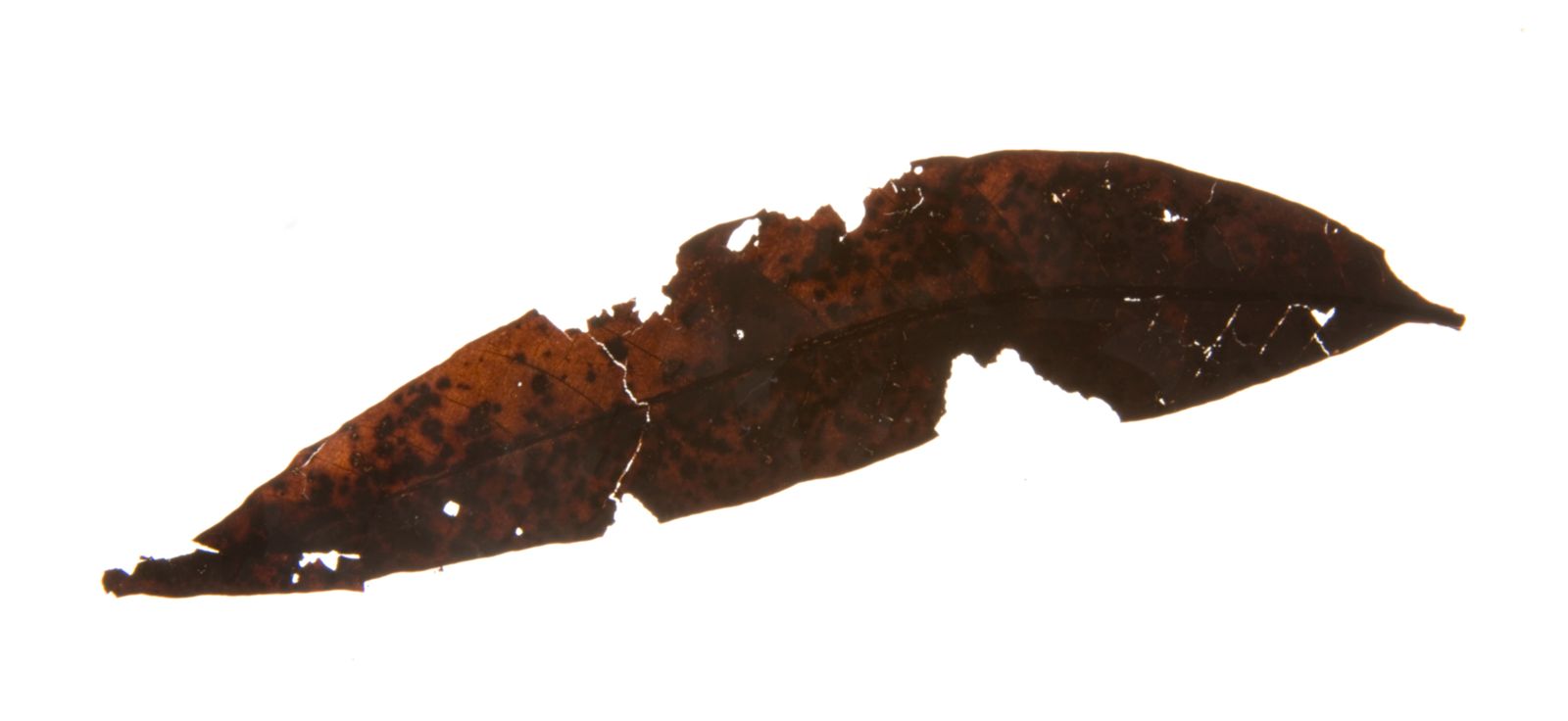
Split the damp clay at Clarkia and, for a few moments, the Miocene can look indecently recent. Field accounts describe leaves emerging in brown, red, or yellow, their blades and veins still distinct, before exposure to air darkens the organic material. The change is fast enough to make excavation part of the spectacle: a leaf that spent roughly 16 million years in oxygen-poor sediment visibly enters a new chemical setting while someone is holding it.[1][9]
That spectacle invites a bad binary. Either the leaf survived, one might think, or it became a conventional stone fossil. Clarkia's leaves did neither. Their outlines, cellular detail, dark organic films, portions of lignin-derived chemistry, and some leaf-wax lipids crossed deep time with very different degrees of alteration. Other compounds were not detected. Famous reports of Miocene plant DNA opened an argument that visual freshness could never settle.[3][4][5][6]
The cover specimen holds the problem in one frame. YPM PB 049087 is recognizably a leaf of Lithocarpus, the stone-oak genus: a midrib runs nearly the length of the blade and smaller veins angle toward its margin. It is also incomplete, removed from its sediment, cataloged with glycerin as a preservation treatment, and photographed long after collection.[11] Those are not blemishes on perfect preservation. They are the preservation story.
Clarkia is therefore best read as a field laboratory for selective survival. The useful question is not, “Is this leaf preserved?” It is, “Which feature survived, in what form, in which layer, and under which test?”
A basalt dam made a lake, not a single frozen afternoon
The fossil beds lie near Clarkia in the Idaho Panhandle, within lake sediments commonly assigned to the Latah Formation. They came to scientific attention in the early 1970s after work on a snowmobile racetrack exposed leaf-rich clay at the locality now called the Fossil Bowl; a University of Idaho account dates that event to 1972. The cut bank beside the track became the type site P-33, one of several Clarkia localities distributed around the footprint of an ancient lake.[2][9]
Columbia River flood-basalt lava blocked the ancestral St. Maries drainage and created that lake. Uranium–lead dating of zircons in interbedded ash now constrains deposition at P-33 to 15.78 ± 0.039 million years ago. The same study identifies annual laminations through about 7.5 metres of section and estimates that interval accumulated over roughly 840 years. That result is more useful than the 17-to-20-million-year estimate carried by some early molecular papers. The change does not make those experiments irrelevant, but it shows why the age printed beside a fossil is itself a geological result subject to revision.[10]
At P-33, an outcrop reported at roughly nine metres thick records more than one event; the newer annual-layer study examined about 7.5 metres of that succession. Clay and silt dominate, with sand low in the section and several volcanic-ash layers higher up. The fine sediment accumulated in a deep lake whose lower water was reconstructed as cold and oxygen-poor. Leaves, needles, fungi, insects, fish, and rare tetrapod remains entered this basin at different moments; Clarkia is an archive assembled by repeated deposition, not one autumn afternoon sealed under a single blanket.[2][9][10]
The lake chemistry did the crucial work. Limited oxygen slowed decay and reduced disturbance on the bottom, while fine sediment covered fragile material. Yet “anoxic” is not a magic word that guarantees uniform fidelity. Conditions differed vertically and through time. Researchers divide the section into a lower, unoxidized zone and an upper, oxidized zone. Fossils occur in both, but the exceptionally detailed plants with preserved organic matter and biomolecular signals come from the lower zone.[2]
That boundary turns the outcrop into a natural comparison. The same broad basin can preserve a leaf-shaped mark above and an organic leaf film below, while only one retains chemistry useful for molecular analysis. Locality and height in the section are therefore part of the specimen, even when the object later sits alone in a museum drawer.
The fresh surface begins changing at the field edge
Clarkia's celebrated color change happens where two preservation regimes meet. Within damp, unoxidized clay, some newly exposed leaves have been described as retaining autumnal browns, reds, and yellows. Once air reaches them, they rapidly darken. The safest interpretation is not that every visible hue is an untouched Miocene pigment. It is that reduced organic material has been protected from modern oxygen, and excavation starts reactions that burial had suppressed.[1][9]
This makes collection more than removal. Time since splitting, moisture, depth, sediment unit, and exposure conditions can affect what an investigator later measures. A photograph taken at the first opening and one made after drying may record different stages of the same specimen's modern history. A chemical sample taken from the lower unoxidized zone and a visually similar imprint from the upper zone should not be treated as interchangeable.
The distinction also sets a limit on the cover image. It documents a genuine Clarkia leaf in a museum collection, with its blade and venation plainly visible.[11] It does not show the leaf's original color, the layer in which it lay, or its chemistry at the instant of exposure. The photograph supports morphology and provenance. Claims about molecules require other instruments.
A leaf survives component by component
In 1993, Graham Logan and colleagues tested Clarkia leaf tissue with pyrolysis mass spectrometry and pyrolysis gas chromatography–mass spectrometry. Their result was a molecular inventory with conspicuous gaps. They detected altered lignin and an aliphatic biopolymer, but did not detect structural polysaccharides, cutin polyesters, or proteins with those methods. A leaf could therefore keep a coherent film and recognizable shape after several major classes of original material had fallen below detection or been transformed.[3]
That does not reduce the dark film to meaningless carbon. Lignin is a structural component of plant tissue, and chemically resistant aliphatic material can retain information about its biological source. But survival is not identity without qualification: a detected product may be an altered residue, and “not detected” means below the method's reach in the tested samples, not metaphysical proof that no fragment exists anywhere in the bed.
Later work pushed the question from “What remains?” to “Can what remains still distinguish plants?” A 2000 study examined 83 fossil leaves from nine genera, including Quercus, Platanus, Magnolia, Fagus, Taxodium, and Metasequoia. The researchers compared distributions of long-chain alkanes and alcohols associated with leaf waxes against the surrounding sediment. Several genera retained repeatable patterns; in many samples, fossil-associated lipids were more concentrated than the matrix background.[4]
That is a genuine form of chemical taxonomy. It is also bounded. Lipid profiles can overlap, migrate, mix with sedimentary organic matter, or change during burial. The method gains force when a chemical pattern repeats across independently identified leaves and differs from adjacent clay. It does not turn one hydrocarbon peak into a species label, much less a complete biochemical portrait.
Clarkia's strongest molecular evidence thus works as a correspondence among three records: visible anatomy, specimen-specific chemistry, and matrix controls. Remove the anatomy and the compound loses its owner. Remove the matrix sample and contamination becomes harder to see. Remove the chemistry and the leaf's fresh look remains only a look.
One squirrel shows how uneven the archive is
The first tetrapod reported from Clarkia makes the same point with bones. Specimen UWBM 113209 was found in 2009 in the lower unoxidized zone at P-33, immediately below an ash correlated with the 15.66 ± 0.07-million-year-old Bully Creek tuff. It preserves part of a skull, forelimbs, vertebral column, ribs, and sternum largely as a carbonaceous film. Only fragments of cheek teeth and part of an incisor remain three-dimensional.[2][10]
Those scraps are enough to constrain, not complete, an identification. The ever-growing incisor establishes a rodent. Low-crowned, rounded tooth fragments, body size, and a comparatively gracile forelimb support placement in the squirrel family, Sciuridae. The authors stopped above genus because the cheek teeth and diagnostic skeletal details are too incomplete. They inferred a tree-climbing habit from the slender limb proportions together with Clarkia's forested environment, while explicitly treating that ecology as comparison rather than a behavior photographed in the rock.[2]
The specimen is a useful counterweight to the leaf abundance. Clarkia did preserve a forest community, but the archive is not a census of that community. Leaves are numerous; fish occur in particular layers; one partial squirrel supplied the site's first reported tetrapod. Different remains reached the lake bottom, decayed, and entered the fossil record with different probabilities. Exceptional preservation can make individual fossils startlingly intimate while leaving the ecosystem statistically lopsided.
DNA has to pass a different test
Clarkia entered molecular-paleontology history in 1990, when a team reported an 820-base-pair PCR product from the chloroplast gene rbcL in a fossil Magnolia leaf and derived a sequence from that product. The work was striking because it proposed information-bearing DNA on a timescale far beyond the ancient sequences then available.[5]
The first complication arrived quickly. In 1991, Arend Sidow, Allan Wilson, and Svante Pääbo analyzed Clarkia extracts, failed to reproduce the reported chloroplast amplification, and concluded that visible high-molecular-mass DNA in some samples was mostly, perhaps exclusively, bacterial. That result exposed a contamination route inside the fossil itself: microbes could contribute abundant DNA even when a plant-shaped film supplied the sample. It did not prove that the original Magnolia product was bacterial, but it did remove bulk DNA yield as evidence that the leaf genome had survived.[6]
The claim did not simply vanish. A 2004 study reported partial chloroplast sequences from Magnolia latahensis and Persea pseudocarolinensis, arguing again for Clarkia's molecular potential.[7] The resulting record is not a clean sequence from announcement to acceptance or rejection. It is a dispute over authentication: whether an amplified molecule originated in Miocene tissue, later microbes, modern plants, laboratory reagents, or a mixture that PCR selectively magnified.
Ancient-DNA authentication keeps separate three propositions that popular retellings often merge. A fossil cell may preserve a stainable or chemically altered DNA-like residue. A sample may contain amplifiable DNA. An authenticated sequence may be shown to belong to the ancient organism. Each step requires different controls. Independent replication, extraction blanks, fragment-length and damage patterns, and comparison with environmental and laboratory sequences all matter; neither recognizable anatomy nor the survival of lignin and lipids can substitute for them.[8]
The responsible conclusion is therefore narrower than either “Clarkia preserved an intact plant genome” or “nothing derived from nucleic acids could remain.” Historical PCR products were attributed to Clarkia plants, but endogenous Miocene DNA has not been securely authenticated; replication failures and contamination concerns mean the sequences are generally treated as artifacts rather than proof of a 15.78-million-year-old plant genome.[5][6][7][8] Clarkia's securely demonstrated lesson is more interesting anyway: one burial setting can preserve morphology, organic films, resistant polymers, and diagnostic lipid distributions while giving every molecular class a different evidentiary ceiling.[3][4]
The fossil is a stack of partial survivals
Return to the dark leaf against white. Its outline supports a plant identification. Its collection record connects it to the Fossil Bowl. Separate analyses of other Clarkia leaves demonstrate that organic matter endured, but this catalog photograph does not reveal its composition. None of those observations alone identifies an ancient protein, pigment, wax molecule, or DNA sequence.[3][11]
That separation is not pedantry; it is what makes Clarkia powerful. The site preserves a mid-Miocene forest near the climatic warmth of the Miocene Climatic Optimum, complete enough to join plant anatomy to insect, fish, fungal, and rare mammal evidence.[2] It also lets researchers watch preservation fail selectively—between layers, between tissues, between compounds, and again when fresh air reaches a newly opened slab.
A time capsule suggests one sealed container with one opening date. Clarkia is closer to a stack of envelopes made from different materials. Some kept their shape. Some kept part of their chemistry. Some lost the message while retaining the paper. The field report becomes accurate only when each envelope is opened on its own terms.
Sources
- Texas A&M University, “Leaf Fossils, What Can You Tell Us From The Past?” (2019)—field observation of newly exposed Clarkia leaf colors and their rapid oxidation, with lake and research context.
- Jonathan J. M. Calede et al., “The first tetrapod from the mid-Miocene Clarkia lagerstätte (Idaho, USA),” PeerJ 6 (2018)—site geology, oxidized and unoxidized zones, a correlated ash, and the partial squirrel UWBM 113209.
- Graham A. Logan, Jaap J. Boon, and Geoffrey Eglinton, “Structural biopolymer preservation in Miocene leaf fossils from the Clarkia site, northern Idaho,” PNAS 90 (1993)—pyrolysis evidence for selective molecular survival.
- Matthew J. Lockheart, Pim F. van Bergen, and Richard P. Evershed, “Chemotaxonomic classification of fossil leaves from the Miocene Clarkia lake deposit, Idaho, USA based on n-alkyl lipid distributions and principal component analyses,” Organic Geochemistry 31 (2000)—leaf-wax lipid comparisons among 83 fossils and surrounding sediment.
- Edward M. Golenberg et al., “Chloroplast DNA sequence from a Miocene Magnolia species,” Nature 344 (1990)—the original report of an amplified Clarkia chloroplast sequence.
- Arend Sidow, Allan C. Wilson, and Svante Pääbo, “Bacterial DNA in Clarkia fossils,” Philosophical Transactions of the Royal Society B 333 (1991)—evidence that high-molecular-mass DNA in Clarkia extracts was predominantly bacterial.
- Sangtae Kim et al., “DNA sequences from Miocene fossils: an ndhF sequence of Magnolia latahensis and an rbcL sequence of Persea pseudocarolinensis,” American Journal of Botany 91 (2004)—later reported chloroplast sequences and their phylogenetic interpretation.
- Svante Pääbo et al., “Genetic analyses from ancient DNA,” Annual Review of Genetics 38 (2004)—critical review of contamination risks and the criteria required to authenticate ancient sequences.
- University of Idaho Library, “Paleobotany & Stratigraphy of Lake Clarkia,” archived 2015 presentation abstract by Bill Rember—1972 discovery, lake reconstruction, anoxic preservation, and reported leaf colors.
- Daianne Höfig et al., “Annually resolved sediments in the classic Clarkia lacustrine deposits (Idaho, USA) during the middle Miocene Climate Optimum,” Geology 49 (2021)—U–Pb age and annual-layer chronology for P-33.
- GBIF and Yale Peabody Museum, “Lithocarpus, YPM PB 049087”—collection record, preservation note, and source for the CC0 fossil-leaf photograph by Linda S. Klise.