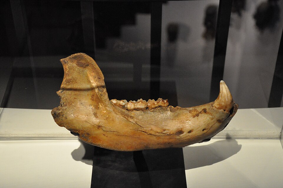
A cave bear’s canine draws attention to its tip: the curve, the point, the damage left by a long working life. Yet a different kind of record survives around the root. Thin layers of cementum, the tissue covering the root, can retain a sequence of growth. Read successfully, that sequence can reveal how long the animal lived and which part of the seasonal cycle was underway when it died.[9]
Those are unusually intimate questions to ask of a fossil. Geological dating places a tooth in deep time; cementum analysis seeks time within the life that produced it. The challenge is to establish that the visible bands preserve the biological rhythm we think they do.
How a root acquires a record
Cementum is part of the tooth’s attachment apparatus. In the acellular tissue used for many age studies, embedded fibers connect the root to the periodontal ligament. As new tissue accumulates around the root, successive layers can preserve approximately annual changes in growth.[4]
The bands have a physical basis. In a 1993 experiment, Daniel Lieberman examined goats given controlled diets. Microscopic differences in their cementum reflected changes in collagen-fiber orientation associated with chewing forces, while differences in mineralization reflected tissue growth. A stripe therefore records properties of the tissue, rather than a date stamped onto it.[2]
That mechanism explains both the attraction and the difficulty. If feeding, growth and seasonal conditions recur regularly, the resulting pattern can become a calendar. But its interpretation depends on understanding the animal and the tissue. A conspicuous line is a starting observation; calling it an annual boundary is a biological claim that needs testing.
Counting requires a starting point
In many mammalian cementum studies, a relatively broad growth zone and a narrower slow-growth band together represent an annual cycle. Counting every visible light and dark band as a separate year would misread that paired structure. Estimating age also requires a developmental correction: the animal had already lived for some time before the relevant tooth began recording its increments.[3]
Researchers can expose the sequence in physical thin sections. Synchrotron X-ray tomography offers another route, resolving internal layers in a three-dimensional dataset. The latter allows a reader to follow a suspected boundary through different views, rather than depending entirely on one physical slice.[9]
Neither instrument supplies the annual interpretation automatically. In their 2024 study of Jurassic mammal growth, Elis Newham and colleagues grounded that interpretation in work on living mammals of known age and comparisons with growth marks in fossil jaw bone. They also screened aggressively for preservation: of 582 imaged specimens, 219 were suitable for counting cementum increments.[4]
That reduction is part of the result. A fossil can retain a recognizable tooth while losing the fine structure needed to read its years. Repeated measurements of damaged tissue may agree with one another without recovering the missing record. More impressive imaging cannot restore a layer that is no longer there.
The outer edge asks a different question
Age estimation reads the accumulated sequence. Seasonal estimation concentrates on its ending: was the last growth zone just beginning, well developed, or followed by a slow-growth band? This demands preservation of the original outer margin as well as a credible seasonal interpretation.[3]
The calendar also varies geographically. A 2019 study of ungulate teeth from Covalejos Cave in northern Spain noted that modern red deer form their slow-growth bands at different times in different European regions. The researchers therefore treated precise ancient calendar assignments cautiously. A tissue category such as active or slow growth does not carry a universal month label.[3]
Cave bears show why even broad seasonal categories matter. Natalya Prilepskaya and colleagues studied twelve fossil canines from European Russia, the Urals and the Caucasus for a paper published in 2022. Two observers independently estimated age and season from cementum; secondary dentine provided another check. They accepted seasonal assignments for ten teeth; disagreement excluded the other two. Six indicated warm-season deaths and four cold-season deaths.[5]
The accepted readings challenged an exclusively winter-denning explanation for those remains. Some bears apparently died in caves during warmer conditions too. But ten seasonal estimates from a selected group of canines cannot establish how frequently every cave-bear population used caves throughout the year. The authors called for further study of individual caves.[5]
The useful change is in the question. Instead of treating a cave full of bear bones as a self-explanatory winter scene, researchers can test when particular animals entered the death assemblage. The tooth narrows the possibilities without furnishing a complete account of the death.
Fourteen years inside a Jurassic tooth
The same method reaches far beyond Ice Age caves. In 2020, Newham and colleagues examined teeth of the Early Jurassic mammal relatives Morganucodon and Kuehneotherium from Welsh fissure deposits, approximately 200 million years old. Their readable sample comprised 34 and 27 specimens respectively, with increments assessed independently by three observers.[6]
The highest counts supported lifespans of at least fourteen years for Morganucodon and nine for Kuehneotherium. These are estimates of the longest lives represented in the sample. They are not average life expectancies, and the oldest recovered individual need not mark a species’ absolute limit. The study also noted that damaged outer increments could make estimates too low.[6]
The authors used longevity to infer aspects of metabolism, prompting a published disagreement. Shai Meiri and Eran Levin argued that body size and the longevity of highly metabolic animals complicate that relationship. Newham’s team defended its comparisons among wild terrestrial species.[7][8] The distinction remains essential: a growth count can inform a physiological argument without directly measuring metabolic rate.
A tooth root thus preserves several possible scales of explanation. Its layers may support an age; its final edge may support a season; comparisons among animals may support a broader account of life history. Each step adds assumptions. The achievement is that, with those steps kept visible, a narrow rim of fossil tissue can bring an individual life into focus.
Sources
- Philippe Alès, “Cave bear jaw, France (Dordogne)” (2015), Wikimedia Commons—photograph provenance and CC BY-SA 4.0 license.
- Daniel E. Lieberman, “Life history variables preserved in dental cementum microstructure,” Science (1993)—controlled-diet goat experiment and the physical basis of banding; NCBI abstract record.
- Carlos Sánchez-Hernández and colleagues, “Combined dental wear and cementum analyses in ungulates reveal the seasonality of Neanderthal occupations in Covalejos Cave (Northern Iberia),” Scientific Reports (2019)—paired increments, developmental offsets, preparation and regional seasonality.
- Elis Newham and colleagues, “The origins of mammal growth patterns during the Jurassic mammalian radiation,” Science Advances (2024)—cementum anatomy, annual-periodicity checks and preservation screening.
- Natalya E. Prilepskaya, Olga P. Bachura and Gennady F. Baryshnikov, “Season-of-death and age-at-death of the easternmost European cave bears,” Boreas (2022)—canine sample, independent readings, seasonal assignments and ecological limits.
- Elis Newham and colleagues, “Reptile-like physiology in Early Jurassic stem-mammals,” Nature Communications (2020)—Welsh fossils, synchrotron imaging, lifespan estimates and potential loss of outer increments.
- Shai Meiri and Eran Levin, “Revisiting life history and morphological proxies for early mammaliaform metabolic rates,” Nature Communications (2022)—critique of the longevity-to-metabolism inference.
- Elis Newham and colleagues, “Reply to: Revisiting life history and morphological proxies for early mammaliaform metabolic rates,” Nature Communications (2022)—defense of the comparative framework.
- Elis Newham and colleagues, “Synchrotron radiation-based X-ray tomography reveals life history in primate cementum incrementation,” Journal of the Royal Society Interface (2020)—growth rhythms and the difference between two-dimensional sections and three-dimensional imaging.