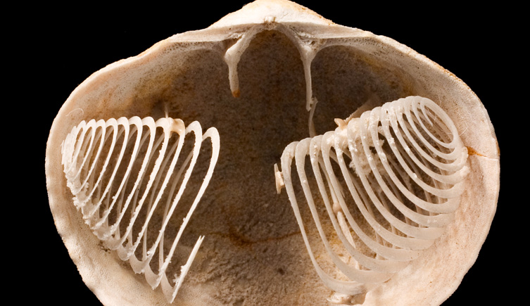
A pair of coils occupies the space where an animal once fed. In the Natural History Museum’s photograph of a Jurassic Spiriferina, the shell’s interior is exposed, revealing a delicate structure called the brachidium. It supported the lophophore, the animal’s soft feeding organ.[1] The fossil offers an unusually intimate view: an internal arrangement that a closed shell would conceal.
The coils invite a satisfying mental reconstruction. Put flesh back along their curves, admit seawater, and the animal seems almost ready to eat. Each step, however, asks a different question. What survives? How can its shape be recovered? And how much of a working animal does that shape actually specify?
A place for the feeding organ
Brachiopods are marine animals enclosed by two shell valves. Their resemblance to clams can be misleading: they belong to a separate animal phylum. They feed with a lophophore, whose tiny beating cilia help move water through the shell, bringing food particles and oxygen. Some brachiopods reinforce this organ with mineral supports.[2]
In the spire-bearing groups, those supports include paired coils called spiralia. The coils extend from structures attached to the dorsal valve, occupying the cavity between the valves.[3] Imagine following one slender strip around its successive turns. The spaces between them matter as much as the strip itself: a reconstruction must leave room for tissue and for water to circulate.
The photograph records the support. Restoring the tentacles and their activity requires comparison with living animals. Keeping those two kinds of evidence distinct lets the fossil remain useful even when researchers disagree about the details of its operation.
Opening the shell without losing the answer
A durable outer shell does not guarantee a durable interior. Coarse sediment entering after death, followed by mineral changes during burial, can damage or destroy the fragile brachidium. Zhiwei Yuan and colleagues emphasized this preservation problem in their study of spiriferide internal anatomy, published online in 2020 and in a 2021 journal issue.[3]
Traditionally, researchers have worked through a fossil in successive physical sections. That exposes structures hidden inside, but consumes the specimen. X-ray microtomography offers a way to recover virtual sections while leaving the object intact.[4] A sequence of slices can then be read as a continuous structure, much as repeated cuts through a coil reveal how its separate-looking cross sections connect.
The scanner still needs something to distinguish. In 2017, Sangmin Lee and colleagues tested 11 Paleozoic brachiopod specimens with different combinations of shell and infilling rock. Original carbonate shells filled largely with quartz grains gave especially clear results. Mineral alteration could reduce the contrast, and unrelated shell fragments in the infill could be mistaken for parts of the brachiopod.[4]
A blurred interior therefore demands restraint. Failure to resolve a coil cannot, by itself, show that the living animal lacked one. Before interpreting anatomy, the researcher has to establish what the specimen and scanning conditions allow them to see.
The direction of dinner
Even a beautifully recovered pair of spirals leaves a problem: which way did water pass through the feeding apparatus?
In 1960, Martin Rudwick reconstructed spiral lophophores by comparison with living brachiopods. His account proposed water entering the shell cavity at the sides and leaving centrally. Within that overall pattern, he inferred different directions of filtration through the spiral cones in forms such as Atrypa and Spirifer.[5]
The important distinction is between a preserved curve and an inferred current. Water has no fossilized arrowhead. Its proposed route depends on how tentacles are restored around the support and how incoming water is separated from water already filtered. A reconstruction can look anatomically persuasive while still needing a mechanical test.
A later approach tested how the shell could harness ambient currents for passive feeding. In 2010, Yuta Shiino and Osamu Kuwazuru modeled Paraspirifer bownockeri, a Devonian brachiopod from Ohio’s Silica Shale. They varied the depth of the sulcus, the central depression in one valve, across four shell models. Their simulations generated inflow through the central sulcus opening and spiral circulation inside.[6]
The natural shell form combined relatively slow internal flow with a stable inflow region. Simply deepening the depression did not improve every aspect of performance. Crucially, the researchers omitted the internal muscles and lophophore to isolate the shell’s effects.[6] The experiment shows what shell geometry can do under specified conditions; it cannot supply a complete replay of feeding. Its scope also explains why a result about Paraspirifer should not automatically be transferred to every animal with spiral supports.
Small attachments, large implications
The coils themselves were only part of the anatomical story. Yuan and colleagues compared 65 spiriferide species and distinguished four arrangements of the supporting apparatus. In some, elongated supporting processes suggested that the mouth may have shifted forward. That is a proposed change in soft anatomy inferred from mineral architecture, rather than a preserved mouth.[3]
This is where the opened shell becomes especially rewarding. Follow the spirals back toward their attachments, and a seemingly decorative structure turns into a set of constraints on where the animal could fit its organs. Follow the spaces outward, and the same structure raises questions about moving water.
The museum specimen preserves the extraordinary delicacy of that arrangement. Its missing soft parts leave work to do: observing living relatives, recovering other interiors and testing how particular shapes behave. The coils give those investigations a physical place to begin.
Sources
- Natural History Museum, London, “Brachiopod collection” — Jurassic Spiriferina photograph and identification of its preserved brachidium.
- Jaleigh Q. Pier, “Brachiopoda.” Digital Encyclopedia of Ancient Life, Paleontological Research Institution (2019) — shell anatomy, lophophore and feeding.
- Zhiwei Yuan, Wen Guo, Dan Lyu and Yuanlin Sun, “Brachial supporting structure of Spiriferida (Brachiopoda).” Journal of Paleontology 95 (2021), 553–567; online 2020 — preservation, support types and inferred mouth position.
- Sangmin Lee and colleagues, “Virtual palaeontology: the effects of mineral composition and texture…” Palaeontologia Electronica 20.2.3T (2017) — X-ray microtomography of brachiopods, mineral contrast and misleading inclusions.
- M. J. S. Rudwick, “The Feeding Mechanisms of Spire-Bearing Fossil Brachiopods.” Geological Magazine 97 (1960), 369–383 — abstract describing the comparative reconstruction and proposed currents.
- Yuta Shiino and Osamu Kuwazuru, “Functional adaptation of spiriferide brachiopod morphology.” Journal of Evolutionary Biology 23 (2010), 1547–1557 — shell-flow models and omission of internal soft parts.