Zika was never only a mosquito story. It looked like one at first because the obvious route was a daytime-biting Aedes mosquito and the usual illness was often mild: fever, rash, conjunctivitis, headache, joint pain, and muscle pain, if symptoms appeared at all.[1][2] That familiar arbovirus frame broke down in 2015 and 2016, when the outbreak in the Americas forced public health to treat a short rash illness as a pregnancy, fetal-brain, and surveillance problem.
The mechanism is a sequence, not a slogan. Zika first has to reach a person, usually through Aedes aegypti in places where the vector can sustain transmission. It can then move through sex or from a pregnant person to the fetus. Once fetal exposure occurs, the most consequential damage clusters around the developing brain and eye. The hard part for public health is that symptoms are too weak a filter: many infections are asymptomatic, and CDC registry data found birth-defect frequencies in exposed pregnancies that were not confined to symptomatic infections.[3][4]
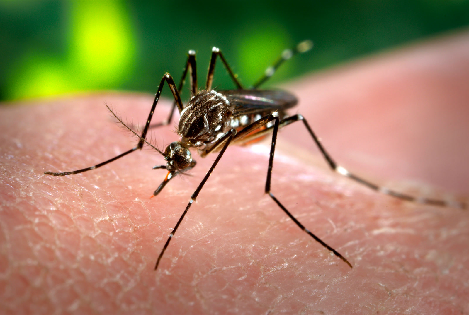
Image context: the cover photograph shows the real mosquito vector rather than an abstract medical symbol. It is useful here because Zika's story begins in vector ecology, then becomes much less visible once the relevant damage depends on pregnancy timing, fetal tissues, and later infant evaluation.[7]
Timeline anchors
- 1947: Zika virus was first identified in Uganda in a rhesus macaque; evidence of human infection and disease followed in African countries in the 1950s.[1]
- 2007 onward: outbreaks were recorded across Africa, the Americas, Asia, and the Pacific, turning an obscure virus into a recurring arboviral threat.[1]
- 2015: Brazil's large epidemic made the association with microcephaly impossible to dismiss as background noise.[1]
- February-November 2016: WHO declared a Zika-related microcephaly and neurologic-disorder Public Health Emergency of International Concern, then lifted it after the causal link between Zika virus and congenital malformations had been confirmed.[1][5]
- 2017 onward: global reported disease declined, but WHO says transmission persists at low levels in several countries, and 92 countries and territories have reported evidence of mosquito-transmitted Zika virus infection.[1]
The first gate is exposure, not illness
Zika's entry point is often ordinary enough to be missed. CDC says the virus is spread mostly by the bite of an infected Aedes species mosquito, with Aedes aegypti as the primary mosquito that spreads Zika.[2] WHO adds that Aedes mosquitoes usually bite during the day, often outdoors but also indoors, and that the vector ecology overlaps with dengue, chikungunya, and urban yellow fever.[1]
That overlap matters because the first symptoms do not reliably identify the virus. WHO gives a typical symptom-onset window of 3-14 days after infection and says symptoms, when present, usually last 2-7 days.[1] Those symptoms are not distinctive enough to carry the diagnosis by themselves. Laboratory confirmation can be complicated by the timing of viral RNA detection and by antibody cross-reactivity with related flaviviruses such as dengue.[1]
The public-health trap is that exposure can matter even when illness barely registers. CDC's public guidance is blunt: many infected people have no symptoms or only mild symptoms, yet the virus can spread through sex and can pass to a fetus during pregnancy.[2] That is why Zika cannot be managed as a simple "feel sick, get tested" disease in pregnancy. The consequential exposure can be quiet.
The second gate is the placenta
The pregnancy boundary is what changed Zika's public meaning. A mosquito-borne infection that might otherwise pass as a brief rash illness became a threat to fetal development because the virus can move from a pregnant person to the fetus.[1][2][3] CDC's congenital-Zika page treats that vertical route as the central fact: infection during pregnancy can lead to congenital Zika syndrome and other birth defects.[3]
The phrase "birth defects" is broad, but the pattern is not vague. CDC lists smaller-than-expected head size, problems with brain development, eye damage, joints with limited range of motion, and excessive muscle tone that restricts movement among conditions that can occur with congenital Zika virus infection.[3] WHO similarly describes microcephaly, limb contractures, high muscle tone, eye abnormalities, and hearing loss as part of congenital Zika syndrome.[1]
The causal claim was not made from one dramatic observation. Rasmussen and colleagues' 2016 review in the New England Journal of Medicine evaluated the evidence for causality at the point when public health had to decide whether Zika was merely associated with birth defects or capable of causing them.[5] WHO's later summary reflects the conclusion that followed: the causal link between Zika virus and congenital malformations was confirmed during the 2016 emergency period.[1]
The third gate is the developing nervous system
Congenital Zika syndrome looks brain-centered because fetal brain development is one of the places where the virus can do disproportionate harm. CDC's registry report emphasizes serious defects of the brain and eyes, including intracranial calcifications, cerebral or cortical atrophy, abnormal cortical patterns, ventriculomegaly, chorioretinal abnormalities, and optic nerve abnormalities.[4]
Laboratory work made that clinical pattern more biologically plausible. A 2016 Cell Stem Cell study reported that a Zika strain efficiently infected human cortical neural progenitor cells derived from induced pluripotent stem cells; infection increased cell death, disrupted cell-cycle progression, and attenuated growth of those progenitor cells.[6] A cell model is not the same as an infant outcome, but it helps explain why the outbreak did not look like a generic congenital infection. The relevant target was not just "the fetus." It was a developmental system where losing cells or slowing growth can alter the architecture of the brain.
That is also why microcephaly became the visible signal but not the whole disease. CDC warns that not every baby with congenital Zika syndrome has all listed findings, that some infants without microcephaly at birth can develop it later, and that some babies who look healthy at birth can develop long-term health problems as they grow.[3] The mechanism can therefore outlast the delivery-room exam.
Timing changes the odds, but it does not create a safe trimester
The U.S. Zika Pregnancy and Infant Registry gives the cleanest numeric frame for the risk boundary. Among 6,799 live-born infants in the registry from pregnancies with laboratory evidence of confirmed or possible Zika virus infection during December 1, 2015-March 31, 2018, 4.6% had any Zika-associated birth defect. In the subgroup with a positive nucleic acid amplification test during pregnancy, the frequency was 6.1%.[4]
Timing mattered. In the NAAT-positive subgroup, the frequency of any Zika-associated birth defect was 8.0% with first-trimester infection, 6.0% with second-trimester infection, and 3.8% with third-trimester infection.[4] That gradient supports the causal mechanism: earlier exposure collides with more foundational stages of organ and brain development. But it does not turn later exposure into a non-event. CDC's report says birth defects were reported with exposures throughout pregnancy.[4]
Symptoms were also a poor gatekeeper. In the same registry, symptoms compatible with Zika were reported in 35% of pregnant women, and birth-defect frequency was similar among infants born to symptomatic and asymptomatic infected pregnant women: 5.3% versus 4.2%.[4] That is one of the most important operational lessons from the outbreak. If surveillance and clinical attention depend too heavily on visible illness, they will miss part of the exposure chain.
Surveillance has to see what the patient may not feel
Zika's decline after 2017 did not erase the mechanism. It changed the surveillance problem. WHO says global cases declined from 2017 onward, but low-level transmission persists in several countries and sporadic outbreaks continue to be documented.[1] CDC's registry authors make a related point from the U.S. outbreak: because symptoms are often mild or absent and testing has a short useful window, combining pregnancy-linked surveillance with birth-defect surveillance was critical to understanding outcomes.[4]
That is the reason Zika remains a useful public-health case study even when headlines fade. The disease taught that a mild adult syndrome can hide a high-consequence fetal pathway; that congenital injury can appear as a pattern of brain, eye, and motor findings rather than a single defect; and that absence of symptoms is not absence of risk in pregnancy.[1][3][4]
The mechanism also sets realistic boundaries. CDC says there is currently no vaccine to prevent Zika and no medicine to treat the disease.[2] Prevention therefore still runs through mosquito-bite reduction, vector control, travel and pregnancy counseling, and attention to sexual transmission, especially where exposure could affect pregnancy.[1][2] None of those measures is glamorous. They matter because they act before the boundary crossing that made Zika historically important.
Zika's lesson is not that every mild virus hides a congenital syndrome. It is narrower and stronger: when a pathogen can move from vector exposure to pregnancy exposure to vulnerable developmental tissue, the apparent severity of the adult illness is the wrong measuring stick. The decisive question is not how sick most infected adults feel. It is whether the virus can reach the fetus at the wrong developmental moment, and whether the health system is built to notice that before the next outbreak makes the answer visible again.
Sources
- World Health Organization, "Zika virus" fact sheet (November 6, 2025) - transmission, symptoms, pregnancy complications, 2016 PHEIC timeline, persistent transmission, and global country count.
- Centers for Disease Control and Prevention, "About Zika" (January 30, 2025) - mosquito, sexual, and pregnancy transmission; common symptoms; and vaccine/treatment status.
- Centers for Disease Control and Prevention, "Congenital Zika Syndrome and Other Birth Defects" (January 31, 2025) - birth-defect pattern, symptom boundary, timing, and future-pregnancy note.
- Roth NM, Reynolds MR, Lewis EL, et al., "Zika-Associated Birth Defects Reported in Pregnancies with Laboratory Evidence of Confirmed or Possible Zika Virus Infection - U.S. Zika Pregnancy and Infant Registry, December 1, 2015-March 31, 2018," MMWR 71, 2022.
- Rasmussen SA, Jamieson DJ, Honein MA, Petersen LR, "Zika Virus and Birth Defects--Reviewing the Evidence for Causality," New England Journal of Medicine, 2016; PubMed record.
- Tang H, Hammack C, Ogden SC, et al., "Zika Virus Infects Human Cortical Neural Progenitors and Attenuates Their Growth," Cell Stem Cell, 2016; PubMed record.
- CDC Public Health Image Library, Image 9258, "female, Aedes aegypti mosquito...acquiring a blood meal from her human host" - real 2006 CDC photograph used as the article image source.