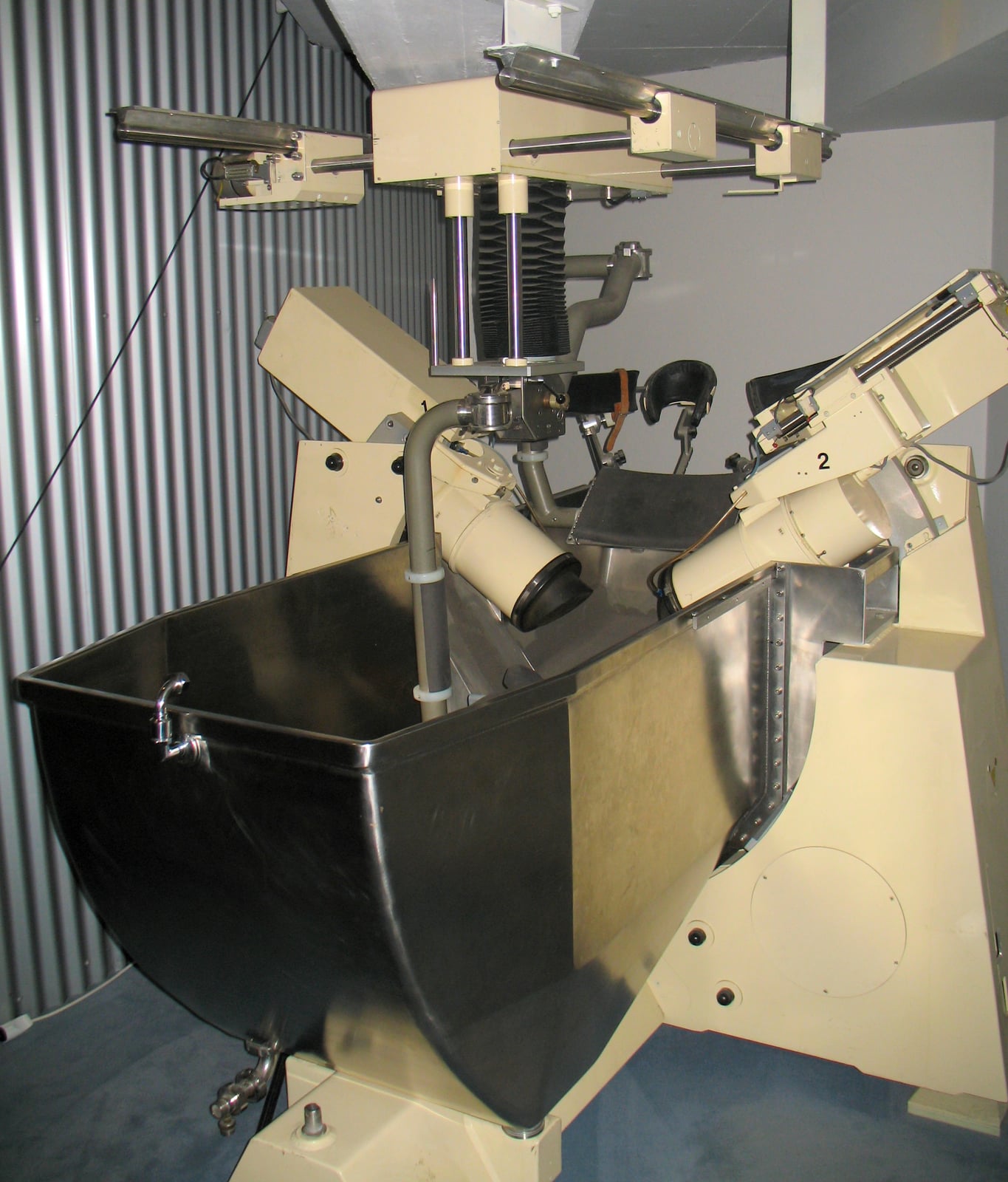
The Dornier HM1 in this museum photograph preserves the imposing equipment behind an unusually delicate promise: reach a kidney stone without making an incision.[1] On 7 February 1980, Christian Chaussy performed the first clinical extracorporeal shock-wave lithotripsy in Munich. Early Dornier treatments required a water bath and general anaesthesia; later machines made outpatient treatment possible.[2]
The bath has largely disappeared from the treatment room, but the procedure still contains two distinct jobs. A machine must turn a stone into smaller pieces. Those pieces must then travel out through the urinary tract. A successful first step does not guarantee a successful second.
That gap explains why a treatment can end on schedule while the patient's stone problem remains unfinished.
Getting the pulse to the stone
A modern appointment makes the procedure sound almost modest. Hull University Teaching Hospitals describes a patient lying on a couch while X-ray or ultrasound imaging locates the stone. Treatment usually lasts 30–40 minutes at that service, with discomfort sometimes resembling repeated tapping against the back. The stone is hidden; the team works through images and carefully positioned equipment.[3]
Between the machine and the body, seemingly minor details matter. A treatment head needs acoustic contact with the skin, usually through a coupling medium such as gel. Air trapped at that interface reflects energy that should be travelling inward.
In a 2006 laboratory study, Yuri Pishchalnikov and colleagues photographed these trapped pockets and measured their effects. Air covering only 2% of the coupling area reduced the breakup of artificial stones by 20–40% under the tested conditions. Separating the head from the test surface and reconnecting it also degraded transmission.[4]
Those are bench measurements, not predictions for an individual patient. Their practical meaning is nevertheless clear: the energy setting on a console cannot tell the whole story of the energy arriving at a stone. Contact is part of treatment, not merely preparation for it.
The 2026 European Association of Urology guideline accordingly calls for careful coupling, repeated imaging checks, and adequate pain relief to limit movement. The target must remain in the intended path while treatment proceeds.[6]
Fracture is a beginning
Once the pulse reaches the stone, more than one physical process contributes to its destruction. Stress waves load the solid material and help fracture it. Cavitation—the growth and collapse of bubbles in the surrounding liquid—contributes to further breakup. Their interaction matters more than a picture of one enormous blow.
Songlin Zhu and colleagues separated these effects experimentally in 2002, exposing artificial stones and calcium oxalate monohydrate stones to shocks in water or castor oil. The different liquids helped distinguish stress-wave effects from cavitation. The combination produced finer fragments than stress-wave action alone; cavitation alone was also less effective than the combined process.[5]
The researchers classified fragments under 2 millimetres as passable. That term needs its laboratory boundary: they measured fragment size, not passage through a living person's urinary tract.[5]
A useful analogy is breaking a large object so it can fit through a doorway. Making the pieces small enough addresses one constraint. It does not move them to the doorway, guide them through a bend, or establish that nothing remains behind. The laboratory endpoint is a necessary part of the clinical story, but it stops before the journey is complete.
The kidney has recesses
The kidney's lower pole makes the distinction visible. Its small collecting spaces, called calyces, are places where fragments can remain after treatment. The EAU guideline states that shock waves can disintegrate lower-pole stones as effectively as stones elsewhere in the kidney, yet clearance is poorer because the fragments often stay in the calyx.[6]
A narrow outlet, a long calyx, or a steep drainage angle can hinder success. Stone size and composition also affect suitability for shock-wave treatment. A clinician choosing a procedure therefore considers both whether the stone is likely to break and whether its remains are likely to escape.[6]
This is why “the stone broke” and “the patient is stone-free” answer different questions. The first describes a change in material. The second describes what is left in a person.
An experiment after the breaking was over
A small 2001 randomized trial by Kenneth Pace and colleagues tested the clearance problem directly. It enrolled 69 patients who still had lower-calyx fragments no larger than 4 millimetres, three months after shock-wave treatment. The patients received either a supervised combination of mechanical percussion, inversion, and increased urine production, or another month of observation.[7]
At the study's one-month assessment, 40% of the intervention group and 3% of the observation group were stone-free. Imaging was assessed by a radiologist unaware of group assignment.[7]
The result is striking because the intervention addressed fragments already present after lithotripsy. It supports the idea that moving fragments is a separate treatment problem. But this was a small, selected population with persistent lower-pole remnants. The combined intervention cannot tell us how much each component contributed, and its percentages are not general success rates for lithotripsy.[7]
The trial examined the journey out after fragmentation had already occurred.
The appointment ends before the process does
Hull's patient information makes room for both kinds of unfinished work: further sessions may be needed to break the stone adequately, or a return visit may check whether it has gone. Fragments can also lodge in the ureter, the tube connecting kidney and bladder, and occasionally require another procedure.[3]
Follow-up belongs inside the treatment's logic. Without it, a team could know that it delivered pulses while remaining uncertain about the outcome that matters. The same leaflet calls for immediate contact with the clinical service for fever, inability to pass urine, severe pain on urination, or worsening bleeding after treatment.[3]
The museum machine records the achievement of reaching a stone from outside the body. The less spectacular achievement comes afterwards: establishing that the pieces have left. Lithotripsy makes its fullest sense when both stages remain in view.
Sources
- Wikimedia Commons, “Nierensteinzertrümmerer HM1.jpg”—photograph of the Dornier HM1 at Deutsches Museum Bonn, uploaded by Journey234 on 30 September 2006; image provenance.
- British Association of Urological Surgeons Virtual Museum, “Managing Stones in the Kidney & Ureter”—first clinical treatment in Munich, early water-bath equipment, and subsequent outpatient development.
- Hull University Teaching Hospitals NHS Trust, “Extracorporeal Shockwave Lithotripsy (ESWL),” updated 30 April 2024—appointment, fragment passage, repeat treatment, complications, and follow-up advice.
- Yuri A. Pishchalnikov and colleagues, “Air Pockets Trapped During Routine Coupling in Dry Head Lithotripsy Can Significantly Decrease the Delivery of Shock Wave Energy,” Journal of Urology, December 2006—laboratory measurements of coupling defects and artificial-stone breakup.
- Songlin Zhu, Franklin H. Cocks, Glenn M. Preminger, and Pei Zhong, “The role of stress waves and cavitation in stone comminution in shock wave lithotripsy,” Ultrasound in Medicine & Biology, May 2002—experimental separation of fracture mechanisms and the fragment-size endpoint.
- European Association of Urology, Guidelines on Urolithiasis, 2026—sections 3.4.5 and 3.4.10.d.2 on treatment delivery, lower-pole anatomy, fragmentation, and clearance.
- Kenneth T. Pace and colleagues, “Mechanical percussion, inversion and diuresis for residual lower pole fragments after shock wave lithotripsy: a prospective, single blind, randomized controlled trial,” Journal of Urology, December 2001—69-patient clearance trial; NLM PubMed record and abstract (XML).