Wilhelm Röntgen is often remembered through one haunted image: a hand, a ring, black space around the fingers, and bones appearing where flesh should have kept its privacy.[1][3] The image deserves its fame, but it can also shrink the historical change. If the story is reduced to one eerie photograph, the real medical threshold becomes harder to see. X-rays changed health care because the winter of 1895-1896 turned a physics discovery into a new diagnostic habit. For the first time, physicians could sometimes inspect a broken bone, a lodged object, or an internal structure without beginning with exploratory cutting or relying only on touch, pain, and educated guesswork.[2][3][4]
That change did not arrive all at once on the night of November 8, 1895. On that evening, according to the Nobel biographical account, Röntgen noticed that a barium platinocyanide screen glowed even though his discharge tube was boxed in black cardboard and the room was dark.[1] He then learned that the new rays passed differently through different materials, and when he held his wife's hand in the path of the rays over a photographic plate, the developed image showed bone shadows, a wedding ring, and the lighter penumbra of soft tissue.[1] The discovery was spectacular. Its medical meaning still had to be built.
What made that meaning durable was speed of conversion. Nature's 1896 account captured the crucial property cleanly: the rays passed with relative freedom through wood and flesh but were stopped more strongly by heavy metals and bone.[3] The National Museum of Health and Medicine's history page gives the practical consequence in modern language: for the first time, interior structures of the human body could be viewed without invasive or exploratory surgery.[2] That sentence is the real hinge. It tells us why the hand image mattered. The image was not only uncanny. It reorganized what counted as preliminary evidence before intervention.
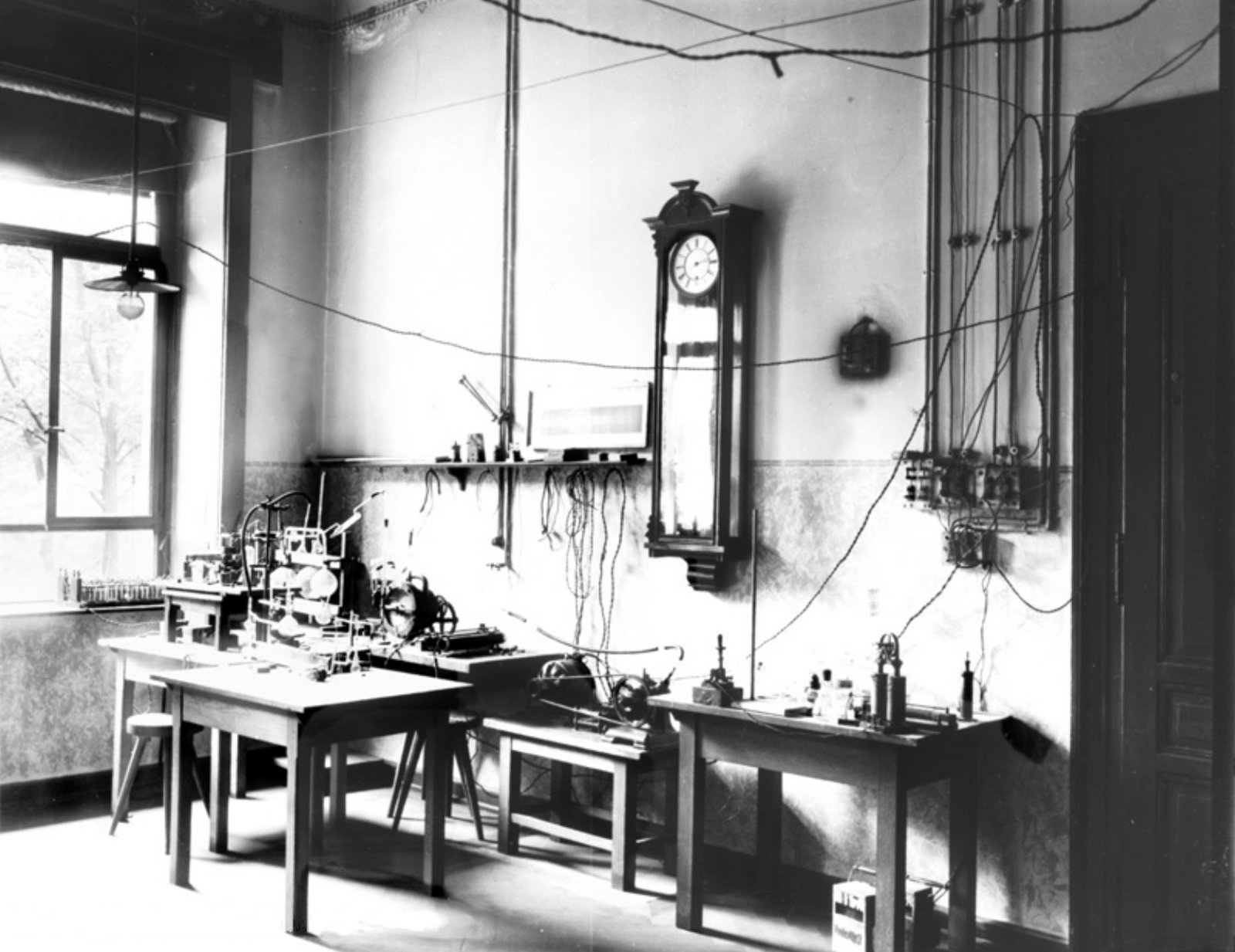
The lead photograph belongs to that story rather than decorating it. It shows the Würzburg room where Röntgen found X-rays, with the discovery still tied to benches, tubes, screens, and a particular working space.[5] That is the right visual because the breakthrough was not "seeing inside the body" in the abstract. It began as a material event in a real room before medicine learned how to turn it into a trustworthy clinical image.
Timeline anchors before the myth hardens
- November 8, 1895: Röntgen observed the fluorescent screen effect in his darkened laboratory and began tracing the behavior of the new rays.[1]
- Late 1895: he made the radiograph of Anna Bertha Röntgen's hand, showing bone shadows and her ring on one plate.[1][2]
- December 28, 1895: Röntgen formally reported the discovery in his paper on a new kind of rays, launching rapid international replication.[3]
- January 1896: news of the discovery spread through Europe and North America, and experimenters began adapting discharge tubes for imaging.[2][3]
- February 3, 1896: Dartmouth's Reed Hall laboratory produced one of the first American clinical X-ray images, examining a patient's broken arm.[4]
These dates matter because they keep the history from collapsing into a single epiphany. The discovery belonged to one night. Medicine changed over the next weeks, when the image stopped being a novelty and started becoming an instrument.
1. Röntgen did not begin with diagnosis; he began with material differences
The laboratory scene on November 8 was not a medical consultation.[1] Röntgen was studying electrical discharge in rarefied gas, building on a line of cathode-ray research already familiar to late nineteenth-century physics.[1] What he found first was not "the inside of the body" but a new relation between radiation and matter. A hidden tube could still excite a screen; interposed substances showed different degrees of transparency; a photographic plate could register those differences.[1][3]
That order matters because it explains why the first medical uses appeared so quickly. The discovery already came packaged as a sorting mechanism. Bone, flesh, wood, and metal no longer behaved the same way under one imaging process.[1][3] A surgeon or physician did not need to master electromagnetic theory to recognize the practical opportunity. If the body could be rendered in layers of different opacity, then hidden anatomy might become visible enough to guide action.
This is also why the hand radiograph became the canonical first image. A hand contains exactly the contrast structure that makes the new ray legible: soft tissue, bone, and metal arranged inside one ordinary body part.[1][2] The ring matters not because it is sentimental, but because it proves comparative density on the same plate. The hand is not a portrait. It is a demonstration that one image can sort the living body into different material classes.
2. The hand image changed medicine because it turned suspicion into inspectable evidence
The famous radiograph of Anna Bertha Röntgen has a theatrical afterlife because it looks like death intruding into domestic life.[1][2] That reading is understandable and incomplete. Its deeper force lies in epistemology. Before X-rays, many internal problems remained inferential. Physicians palpated, listened, measured external deformity, asked about pain, and when necessary cut to find out. After X-rays, at least some hidden structures could be turned into inspectable evidence before the knife entered.[2][3]
The National Museum of Health and Medicine is especially useful here because it states the clinical threshold without melodrama: X-ray technology provided a way to view interior structures without invasive or exploratory surgical procedures.[2] This does not mean medicine immediately became noninvasive in the modern sense. Early radiographs were crude, exposure times were long, and many conditions remained beyond reach. What changed was the sequence of knowing. An internal object no longer had to remain wholly hypothetical until the body was opened.
That is a profound change in medical authority. A physician's judgment could now be supported by a plate that others could inspect. The image was not infallible, but it was shareable. It could be carried, debated, taught, and compared. In that sense, Röntgen's real contribution to health care was not an isolated invention but a new kind of witness.
3. The first winter of X-rays made fracture care newly visible
Dartmouth's 1896 record is valuable because it shows how quickly that new witness entered practice. The library exhibit describes Professor Edwin B. Frost examining a patient's broken arm in Reed Hall and calls it one of the first X-ray experiments in America, joining basic science and medical innovation.[4] That phrasing is precise enough to keep the scale honest. The experiment was still experimental. It was also already clinical.
The fracture example matters because it makes the utility of X-rays immediate. A broken arm had always been partly visible from the outside through pain, swelling, deformity, and loss of function. But those signs did not always tell a physician exactly where the break sat, how fragments aligned, or whether reduction had truly succeeded. Once a plate could show the fracture line itself, the problem changed shape. The body no longer had to speak only through symptoms and manipulation. It could present a shadow record of the injury.[2][4]
That is the winter in which diagnostic medicine shifted. Röntgen discovered rays in Germany. Within weeks, other rooms were using them to ask practical questions about patients.[2][3][4] The transition was startlingly fast because the use case was obvious. Fractures, foreign bodies, and skeletal anatomy already demanded decisions. X-rays did not invent those decisions; they altered the evidence available before making them.
Seen this way, the story is not simply one of faster technology adoption. It is a story about how quickly medicine recognized a new bargain. The exposure might be awkward, the equipment temperamental, and the image quality limited, yet the trade was still worth making because the alternative was greater uncertainty.
4. The larger breakthrough was diagnostic time, not visual wonder alone
It is tempting to say that Röntgen made the body transparent. That phrase is memorable and slightly misleading. Early X-rays did not make everything transparent, and they certainly did not make interpretation effortless.[2][3][5] What they did was create diagnostic time before incision. Physicians could pause, position, expose, develop, and inspect. The image inserted a new stage between first suspicion and operative intervention.
That new stage had consequences well beyond broken bones. It made preoperative planning more concrete. It gave hospital teachers an image they could show students and colleagues. It created records that could outlast the bedside encounter. It also invited overuse and, eventually, taught painful lessons about radiation injury to early operators, who did not yet understand the risks they were taking.[2] The new medical gaze arrived with power and with cost.
Keeping that boundary visible improves the history. Röntgen did not hand medicine a perfect window. He opened a threshold that others then expanded, standardized, and made safer over time.[2][3] The first winter's achievement was narrower and still enormous: some internal truths became photographable.
Why this microhistory still matters
Röntgen still matters in 2026 because modern medicine repeatedly turns on moments when hidden structure becomes inspectable without opening the body. CT, fluoroscopy, mammography, and countless ordinary radiographs all descend from the same historical turn.[2][3] The foundational move was not glamour. It was the conversion of interior anatomy into evidence that could be obtained before exploratory surgery and shared after the fact.
If the hand image is remembered only as a gothic curiosity, the medical lesson disappears. The real change was that diagnosis no longer had to stay outside the body until incision. In the winter of 1895-1896, Röntgen's laboratory discovery crossed into health care and gave physicians a new kind of proof: not certainty, not safety, and not total bodily transparency, but a plate on which flesh, bone, and metal could finally be told apart.[1][2][3][4]
Sources
- Nobel Prize Outreach, "Wilhelm Conrad Röntgen – Biographical" - Nobel biographical account covering the November 8, 1895 discovery, the fluorescent barium platinocyanide screen, and the first hand radiograph of Anna Bertha Röntgen.
- National Museum of Health and Medicine, "Discovery of the X-ray: A New Kind of Invisible Light" - museum history page explaining how X-rays made interior human structures visible without invasive or exploratory surgery.
- Nature, "The Röntgen Rays" (1896) - early scientific account emphasizing the different transparency of flesh, bone, wood, and metal and the rapid public and scientific response to the discovery.
- Dartmouth Library, "Early x-ray experimentation at Dartmouth, 1896" - archival exhibit note on one of the first American clinical X-ray experiments examining a patient's broken arm in Reed Hall.
- Wikimedia Commons, "File:Room where Röntgen found x-rays.jpg" - source page for the archival photograph of the Würzburg room used as the article image.