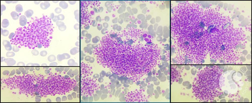
A blood count recorded 23,000 platelets per microlitre. Repeating the test with a different collection tube produced 207,900. In the case published by Oussama Grari and colleagues in the American Society of Hematology's Image Bank on December 30, 2024, the microscope supplied the missing explanation: platelets had gathered into clumps in the first specimen.[1]
The striking change was a correction to the measurement. The case illustrates pseudothrombocytopenia: an apparent shortage of platelets created outside the body. Understanding it requires following the sample past the needle, through the tube's chemistry, and into an instrument that must decide which particles count as platelets.
The tube changes the conditions
EDTA, a common anticoagulant in blood-count tubes, binds calcium and keeps collected blood from clotting. But calcium also helps maintain the structure of proteins on platelet surfaces. In susceptible samples, removing it can expose a site that an antibody can recognize.[2][4]
A March 1986 investigation by H. H. van Vliet and colleagues examined this process in one patient's serum. Antibody binding depended on EDTA concentration and temperature. Their experiments identified platelet glycoprotein IIb as the target and supported the explanation that EDTA made the binding site accessible by altering the calcium-dependent IIb/IIIa complex.[2] The important change occurred in the specimen's environment: the collection method helped create the conditions for the misleading result.
A larger mechanistic study, published by Alessandra Casonato and colleagues in July 1994, investigated 33 patients with pseudothrombocytopenia. Transferring patients' serum or immunoglobulins into normal EDTA-treated blood reproduced clumping. Blocking a relevant site on the IIb/IIIa receptor prevented the effect in their experiments.[3]
That transfer is the revealing step. It showed that something carried in the serum could make otherwise normal platelets behave differently under laboratory conditions. The study supported an antibody-mediated mechanism; it did not establish that every unexpectedly low platelet result has the same cause.
A cluster falls outside the count
A counting instrument does not simply photograph every platelet and tick it off. One widely used method measures changes in electrical impedance as particles pass through a small aperture. The signal reflects particle volume, allowing the analyzer to assign objects within a particular size range to the platelet count.[4]
A clump disrupts that classification. Its constituent platelets are no longer presented as separate platelet-sized objects. Depending on its size and the instrument, the cluster can be excluded from the platelet tally or interfere with another cell count. The blood still contains the platelets; the reported number has lost track of them.[4]
The 2020 recommendations from the French-Speaking Cellular Hematology Group emphasize microscopy when clumping is suspected. Instrument warnings and unusual particle distributions provide clues, but the smear lets a laboratory professional inspect the objects responsible.[4] This is why a technically precise-looking number can require a visual check before it becomes a clinical fact.
The sample also has a clock. In work first published online in November 2014, Jianhua Lin and colleagues followed EDTA and citrate specimens from 26 patients. The artifact could appear within 10 minutes of collection, and weaker clumping also developed in many corresponding citrate specimens.[5] For an affected sample, a falling count during a delay can describe changes inside the tube rather than deterioration inside the patient. Repeating the same measurement later need not provide independent reassurance.
A second tube is another experiment
The Croatian laboratory recommendations published in October 2024 begin by excluding other collection problems. A clot or fibrin strand can make a sample unsuitable; contamination with infusion fluid can dilute cell counts. These possibilities require attention before attributing the result to EDTA-dependent antibodies.[6]
After microscopy confirms clumps, that guidance calls for withholding the unreliable platelet result and communicating with the clinician. Its usual next step is a sodium citrate specimen, with allowance for dilution by the liquid anticoagulant and inspection for persistent clumping.[6] The second tube changes a suspected cause of the error. Merely producing another number does not prove that the cause has disappeared.
There is real disagreement about the best replacement. A GFHC study published online in January 2025 compared EDTA samples containing clumps with paired specimens collected using sodium citrate, CTAD or magnesium sulfate. Its sodium citrate comparison included 335 paired samples. Magnesium sulfate was most effective at avoiding clumps, followed by CTAD; the authors argued that these alternatives should replace sodium citrate for this purpose.[7]
That conclusion goes further than the Croatian pathway. It also explains why the successful citrate result in the opening case cannot become a universal rule. Laboratories need a method that works for the specimen, the available equipment and the way the result is verified. The disagreement concerns how best to recover a trustworthy count, not whether the original clumps can be ignored.[6][7]
Finding the artifact is only part of the answer
A smear showing clumps establishes a problem with that sample. It does not establish the patient's true platelet count. The laboratory still has to obtain a reliable result, and the clinician still has to interpret it alongside the patient's condition.[6]
That boundary gives this small laboratory drama its significance. An unexpected low count deserves investigation; knowing that false lows exist is no reason to dismiss one. Equally, treating a number before checking the specimen can turn an artifact into unnecessary care. The useful question is concrete: were the platelets actually scarce, or had the measurement stopped seeing them separately?
Sources
- Oussama Grari and colleagues, “EDTA-Dependent severe pseudothrombocytopenia.” ASH Image Bank, December 30, 2024. Clinical case and source micrograph.
- H. H. van Vliet, M. C. Kappers-Klunne and J. Abels, “Pseudothrombocytopenia: a cold autoantibody against platelet glycoprotein GP IIb.” British Journal of Haematology, March 1986. Experimental evidence for antibody binding and EDTA-dependent exposure of its target.
- A. Casonato and colleagues, “EDTA dependent pseudothrombocytopenia caused by antibodies against the cytoadhesive receptor of platelet gpIIB-IIIA.” Journal of Clinical Pathology, July 1994. Mechanistic investigation in 33 patients.
- Véronique Baccini and colleagues, “Platelet Counting: Ugly Traps and Good Advice. Proposals from the French-Speaking Cellular Hematology Group (GFHC).” Journal of Clinical Medicine, March 16, 2020. Counting technologies, interference and smear examination.
- Jianhua Lin and colleagues, “Discovery and Correction of Spurious Low Platelet Counts due to EDTA-Dependent Pseudothrombocytopenia.” Journal of Clinical Laboratory Analysis, 2015; first published online November 25, 2014. Time-course experiments in EDTA and citrate specimens.
- Lara Milevoj Kopčinović and colleagues, Croatian national recommendations on management of samples with suspected EDTA-induced pseudothrombocytopenia. Biochemia Medica, October 15, 2024. Collection errors, microscopy, citrate testing and reporting.
- Soraya Wuilleme and colleagues, “Influence of Anticoagulants on Platelet Counts: A Study and Recommendations From the French Speaking Cellular Hematology Group (GFHC).” International Journal of Laboratory Hematology, 2025; first published online January 16. Paired-sample comparisons of alternative anticoagulants.