Obstetric ultrasound is now so ordinary that its hardest historical achievement can disappear behind familiarity. The technology is often remembered as the moment medicine first “saw the baby.” That description catches the emotional afterlife and misses the clinical event.[1][2] Ultrasound became obstetric infrastructure only after it learned to do three narrower jobs in sequence: sort uncertain findings inside the pregnant abdomen, convert fetal size into a more reliable pregnancy clock, and then make fetal abnormality visible early enough to alter management.[2][3][4][5][6]
That sequence matters because the earliest work was not yet the modern anatomy scan, and it was certainly not a keepsake industry. BMUS's historical summary places Ian Donald and his Glasgow colleagues at the practical turning point in the mid-1950s, when ultrasound started moving from physics and industrial technique toward usable medical applications.[1] The classic 1958 paper confirms the original frame in its own title: Investigation of Abdominal Masses by Pulsed Ultrasound.[2] Pregnancy was part of that world, but the first problem was broader and less sentimental. Clinicians needed a way to distinguish what hands, stethoscopes, and plain radiology could not reliably sort inside the pelvis and uterus.
The strongest reading of the years from 1958 to 1973 is therefore not “first image, then inevitable progress.” It is a tighter event reconstruction. One Glasgow-linked breakthrough made ultrasound clinically interesting. A second wave of measurement work made it useful for dating. A third move made it consequential for prenatal decision-making. By the early 1970s, the machine had stopped being a promising curiosity and had started behaving like a timed visual record of pregnancy.[1][2][3][4][5][6]
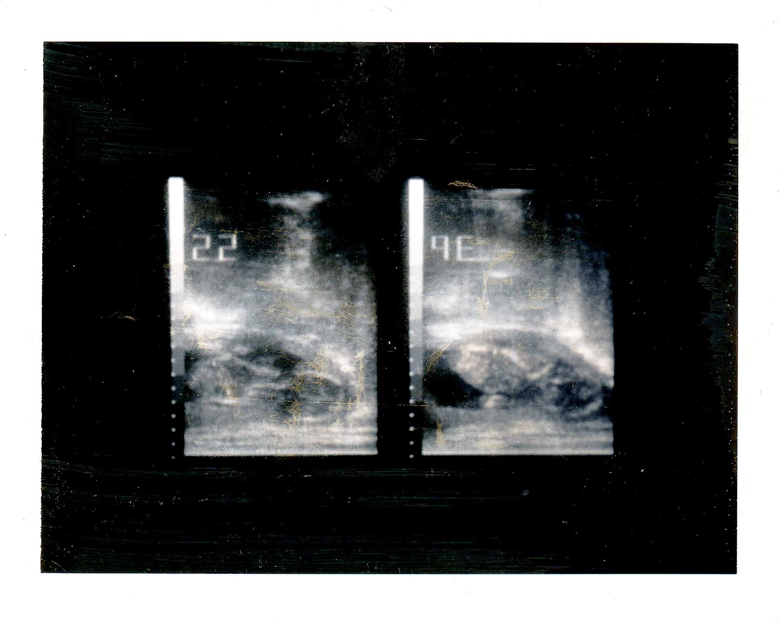
Image context: the lead image is an archival Polaroid photograph of an obstetric ultrasound screen taken in 1985.[7] It belongs here because it shows the mature practical outcome of the earlier decades reconstructed below: a pregnancy scan that could be stored, carried, and compared, rather than a fleeting experiment witnessed only by the operator.
Timeline anchors
- 1958: Donald, MacVicar, and Brown published Investigation of Abdominal Masses by Pulsed Ultrasound, the Glasgow paper that helped move pulsed ultrasound into practical obstetrics and gynecology.[1][2]
- 1966: Goldberg and colleagues published Ultrasonic fetal cephalometry, showing that fetal head measurement by ultrasound could become a clinical method rather than a visual novelty.[3]
- 1971: Whittingham published The ultrasonic biparietal diameter, expressed in time units, reinforcing the idea that one fetal dimension could function as a gestational clock.[4]
- 1972: Campbell and colleagues published Anencephaly: early ultrasonic diagnosis and active management, demonstrating that ultrasound had crossed into prenatal anomaly diagnosis with management consequences.[6]
- 1973: Robinson reported first-trimester crown-rump-length dating from 214 examinations in 80 patients, with pregnancy maturity predicted to within three days between the sixth and 14th weeks.[5]
1. In 1958, the machine entered obstetrics as a sorting tool
The 1958 starting point is revealing because it was still framed around uncertainty rather than reassurance. Donald's Lancet paper was about abdominal masses.[2] That phrasing matters. The early scan room was not yet dominated by routine fetal anatomy review or standardized gestational dating. It was dealing with a harder diagnostic landscape in which fibroids, ovarian cysts, hydramnios, twins, and pregnancy itself could create overlapping physical findings, while ordinary examination remained indirect.[2]
That is why BMUS's historical note is useful. It does not describe Glasgow as a moment of instant perfection. It says Donald and his colleagues helped facilitate practical technology and applications, and that this work led to wider medical use in the following decades.[1] In other words, the first event was not complete adoption. It was technical credibility. Ultrasound had to become something clinicians could act on before it could become something departments organized around.
Seen this way, the earliest obstetric contribution was epistemic. Ultrasound let the examiner replace some inference with a patterned echo response.[2] The fetus, the uterus, and the mass inside the pelvis were no longer entirely matters of touch, symptoms, and delayed confirmation. A new visual discipline began entering the chart, even if the images were static, technically awkward, and still far from the clean grayscale scans later generations would treat as ordinary.
2. In the late 1960s, ultrasound became a clock
Clinical interest alone was not enough to make the machine routine. A routine obstetric technology has to answer a repeated question cheaply and clearly, and one of the most repeated questions in pregnancy is simple: how far along is this pregnancy, really?
That is where fetal measurement changed the status of ultrasound. Goldberg's 1966 paper on ultrasonic fetal cephalometry showed that fetal head dimensions could be treated as measurable clinical data rather than as mere visual impressions.[3] Whittingham's 1971 paper pushed the point further in its own title by expressing the biparietal diameter in time units.[4] Once a fetal dimension could be translated into gestational age, ultrasound stopped being only a problem-solving tool for unusual cases. It started becoming a way to discipline the calendar of ordinary care.
That shift should not be understated. Menstrual recall is often imperfect. Fundal height and quickening are useful but imprecise. A machine that could estimate maturity from fetal size offered a new kind of obstetric authority: one less dependent on memory, body habitus, or late confirmation at delivery.[3][4] Ultrasound gained institutional force when it attached itself to scheduling, induction timing, growth expectations, and the expected date of confinement.
The 1973 Robinson paper shows how sharp that transition had become by the early first trimester.[5] Robinson reported a sonar method for measuring fetal crown-rump length, built a normal curve from 214 examinations on 80 patients, and in a later blind series found that maturity could be predicted to within three days between the sixth and 14th weeks.[5] That is a stronger historical milestone than a generic claim that “ultrasound improved dating.” It marks the point when pregnancy time itself became more formally machine-readable.
This was the real administrative revolution. Once gestation could be dated more accurately, every later comparison became cleaner. A fetus that looked small late in pregnancy might represent wrong dates or true growth restriction; better early dating helped separate those possibilities. A scan machine that measures time well does not merely create images. It restructures what later images mean.[4][5]
3. By 1972, ultrasound had crossed into management
Dating alone would have made ultrasound useful. It would not yet have made it ethically and clinically transformative. That third turn appears when anatomy becomes legible enough to redirect a pregnancy's course.
Campbell and colleagues' 1972 paper, Anencephaly: early ultrasonic diagnosis and active management, is therefore a hinge document.[6] Even before one opens the details, the title shows that ultrasound had moved into a different clinical register. The scan was no longer only helping the clinician estimate maturity or classify a pelvic problem. It was identifying a catastrophic fetal abnormality early enough that management decisions followed from the image.[6]
This is the moment when ultrasound stopped behaving like an adjunct and started behaving like a decision instrument. A dated pregnancy can still remain largely private inside the exam room. A structurally abnormal pregnancy changes counseling, follow-up, and sometimes whether the pregnancy continues.[6] The machine had crossed from chronology into consequence.
That is why the years around 1972-1973 deserve to be treated as a coherent event rather than as isolated technical papers. One line of work made fetal size measurable in time.[4][5] Another line made fetal abnormality diagnosable before birth.[6] Together they turned ultrasound into a way of seeing pregnancy as a sequence of inspectable, comparable, and actionable states.
4. Why the dated image mattered more than the first picture
Public memory often prefers the first recognizable fetal image because it is emotionally legible. The stronger historical argument is colder. Obstetric ultrasound became routine when it gave clinicians a dated image they could compare across visits and interpret inside management pathways.[3][4][5][6] The dated image did more work than the first picture.
This also explains why the archival 1985 Polaroid used here feels historically apt.[7] It shows ultrasound after the pioneering phase, when an obstetric image was expected to be documented, stored, and shown beyond the machine itself. That physical trace matters. A stored scan can be reviewed, measured, argued over, and entered into a pregnancy record. It belongs to the bureaucracy of care as much as to the theater of pregnancy.
The dated image changed the social experience of pregnancy too, but that afterlife followed the clinical ordering work, not the other way around. First the machine had to prove it could sort uncertainty. Then it had to prove it could keep time. Then it had to prove it could disclose anatomy with enough reliability that clinicians would reorganize decisions around it.[2][3][4][5][6] Emotional attachment, souvenir prints, and public familiarity came later, built on top of that older clinical sequence.
What the reconstruction shows
The history of obstetric ultrasound is easiest to flatten into a single miracle of visibility. The more accurate reconstruction is staged. Glasgow made the technique practically credible in 1958.[1][2] Cephalometry and biparietal dating made it administratively valuable in the late 1960s and early 1970s.[3][4] First-trimester crown-rump-length work made gestational timing much tighter by 1973.[5] Prenatal diagnosis of anencephaly showed that the image could now alter management itself.[6]
That sequence is why ultrasound stayed. A machine survives in medicine when it answers recurrent questions better than the older toolkit. Obstetric ultrasound did that by turning pregnancy into something that could be seen, measured, dated, and compared inside one visual record. The first image mattered. The dated image changed practice.
Sources
- British Medical Ultrasound Society, "The History of Ultrasound" - BMUS overview noting that Ian Donald and his Glasgow colleagues in the mid-1950s helped facilitate practical ultrasound technology and that their work led to wider medical use in the following decades.
- Ian Donald, John MacVicar, and Tom Brown, Investigation of Abdominal Masses by Pulsed Ultrasound (Lancet, 1958) - BMUS-hosted copy of the classic Glasgow paper that brought pulsed ultrasound into practical obstetrics and gynecology.
- Bernard B. Goldberg, H. Jack Isard, Joshua Gershon-Cohen, and Bernard J. Ostrum, "Ultrasonic fetal cephalometry" (Radiology, 1966) - early paper establishing fetal head measurement by ultrasound as a clinical method.
- T. A. Whittingham, "The ultrasonic biparietal diameter, expressed in time units" (British Journal of Radiology, 1971) - paper showing how a fetal dimension could function as a gestational-age measure.
- H. P. Robinson, "Sonar measurement of fetal crown-rump length as means of assessing maturity in first trimester of pregnancy" (British Medical Journal, 1973) - first-trimester crown-rump-length method derived from 214 examinations in 80 patients, with maturity predicted within three days between the sixth and 14th weeks.
- S. Campbell, F. D. Johnstone, E. M. Holt, and P. May, "Anencephaly: early ultrasonic diagnosis and active management" (Lancet, 1972) - early paper marking ultrasound's move into prenatal anomaly diagnosis with direct management consequences.
- Wikimedia Commons, "File:Obstetric Ultrasound Polaroid Photograph 1985.jpg" - source page for the archival image used as the article cover.