A small piece of tissue can change the course of an operation. If cancer cells reach the edge of a removed specimen, the surgeon may consider taking more tissue. If a suspicious mass proves benign, the operation may be more limited. Either finding is most useful while the team can still act on it.[2]
The frozen section brings microscopic examination into that interval. Its central trick is physical: freezing makes soft tissue firm enough to slice before the slower preparation of a permanent specimen is complete.[1] That saves time, but leaves two questions intact: was the right tissue selected, and is the resulting view clear enough to support the decision?
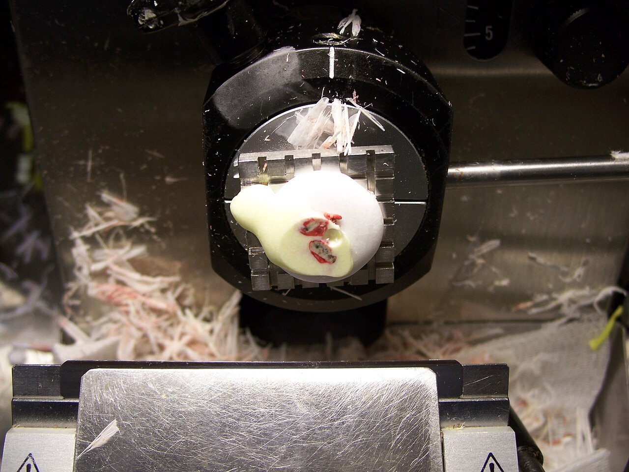
Making soft tissue sliceable
Microscopy requires a thin section through the specimen. In routine preparation, formalin stabilizes tissue, water is removed, and paraffin wax provides support for cutting. The resulting sections preserve fine structural detail, but the processing takes time.[1]
For an intraoperative consultation, fresh tissue goes directly from the operating room to pathology. The pathologist first examines it without magnification and selects the area to investigate. The chosen sample is frozen in a supporting medium, sliced, transferred to a slide and stained. Dyes make the cellular structures visible under the microscope.[2]
The cryostat is the instrument used to cut the frozen sample. The National Cancer Institute describes a frozen-section examination taking about 15–20 minutes. Its explanation also emphasizes that permanent sections usually preserve more detail.[1] The speed comes from a different preparation method; it does not mean that every part of the eventual pathology investigation has been compressed into those minutes.
This tension was explicit in Louis B. Wilson's paper published on December 2, 1905. Working at the Mayo laboratories, he described earlier methods as either too slow for a patient waiting under anesthesia or insufficiently clear. His answer combined fresh tissue frozen in dextrin with sections 10–15 micrometers thick and rapid staining.[5]
What remains recognizable in Wilson's report is the practical problem: preserve enough visible structure to make a useful judgment before the operation moves beyond it.[5]
The question travels with the specimen
A rapid slide needs a specific purpose. The 2021 specimen-handling guidelines from the College of Pathologists of Sri Lanka say that frozen section should be used when its result can influence the surgical procedure. They also call for discussion between clinician and pathologist about the examination's purpose and limitations.[4]
That conversation determines what counts as a useful sample. A question about a mass and a question about its cut edge direct attention to different places. When margins are being assessed, the specimen needs orientation and identification of the relevant edges. Otherwise, a microscopic finding becomes difficult to translate back into the operation.[4]
Imagine a report that cancer cells are present at an edge, but no one can connect that edge to its location in the patient. The cells have been recognized; the surgical question remains unresolved. This is why labeling and orientation belong to the diagnostic process itself.
The decision also has consequences beyond the slide. As the American Cancer Society explains, removing additional tissue can help address an involved margin, but it carries risks that the surgeon must weigh. A pathology finding informs that choice; it does not settle the balance of benefits and complications by itself.[2]
What cold cannot solve
Freezing can distort the structures being examined. Auckland Hospital's frozen-section test guide identifies ice-crystal artifact as one reason microscopic detail may be less clear. It separately identifies sampling as a limitation: small areas of disease can fall outside the sections examined.[6]
These are different failures of information. In one, the relevant tissue is on the slide but its appearance is altered. In the other, the slide may be beautifully readable while the important focus is elsewhere. Improving image quality cannot reveal cells that were never sampled.
More sampling has a cost in time. The Sri Lankan guidelines note that multiple biopsies take longer to process and interpret. They also identify fatty or fibrous tissue as difficult to section.[4] The material arriving at the laboratory therefore helps determine how quickly a useful answer is possible.
Auckland lists 30 minutes from laboratory receipt for a case requiring one block, with complexity affecting turnaround.[6] That local service expectation and the NCI's general estimate are not interchangeable stopwatches: they describe different scopes. A patient or family should not read a longer wait as a diagnosis in itself.
Sometimes the appropriate answer is to defer the diagnosis until permanent sections are available. Auckland explicitly describes this option when frozen tissue cannot support a sufficiently secure interpretation.[6] Deferral preserves uncertainty in the report instead of turning the pressure for speed into false confidence.
The examination continues after the call
The rapid interpretation leaves a record that can be checked. The College of American Pathologists' March 2025 specimen-handling guide calls for retaining intraoperative slides and processing the residual examined tissue into paraffin for comparison with the frozen-section interpretation. It also places the intraoperative consultation in the final surgical pathology report.[7]
Communication is part of that continuity. CAP specifies direct verbal reporting to the intraoperative medical or surgical team, with the patient's identity confirmed before the result is delivered.[7] The useful endpoint is an understood finding attached to the correct patient and specimen.
The resulting chain connects two moments of care. During surgery, the team needs an answer narrow enough to guide an immediate choice. Afterward, pathology can complete the examination with the detail and additional work the case requires. The frozen section earns its speed by answering a particular question—and by making clear when that question must remain open.
Sources
- National Cancer Institute, “Pathology Reports” — frozen and permanent tissue preparation, microscopy and the approximate rapid-examination interval.
- American Cancer Society, “How Is a Biopsy Done?,” revised March 24, 2026 — specimen selection, frozen-section workflow and surgical margin decisions.
- 000jaw, “Tissue for frozen section in cryostat.JPG,” February 17, 2010, Wikimedia Commons — original photograph, description and CC BY 3.0 attribution.
- College of Pathologists of Sri Lanka, National Guidelines in Histopathology: Collection, Handling and Transport of Surgical Specimens, second edition, 2021, printed pages 45–47 — indications, orientation, sampling and technical limitations.
- Louis B. Wilson, “A Method for the Rapid Preparation of Fresh Tissues for the Microscope,” JAMA 45(23):1737, December 2, 1905 — original account of rapid freezing, sectioning and staining.
- Northern Region Pathology Network, Auckland Hospital, “Frozen Section (Fresh Tissue),” updated June 15, 2026 — service turnaround, freezing artifact, sampling limitations and deferred diagnosis.
- College of American Pathologists, Practical Guide to Specimen Handling in Surgical Pathology, version 12.0, March 2025, pages 23–25 — intraoperative reporting, slide retention and comparison with paraffin sections.