On the morning of May 14, 1993, a 19-year-old competitive runner became desperately short of breath while traveling from Crownpoint to Gallup, New Mexico. His family stopped at a service station to call for help. By the time the ambulance arrived, he had collapsed; despite resuscitation at Gallup Indian Medical Center, he died with his lungs filling with fluid. His fiancée, who shared his home, had died after a similarly abrupt illness only days earlier.[2][3]
The scene looked like a medical impossibility: two young, active Diné people, a brief fever-and-muscle-ache illness, then overwhelming pulmonary edema. Yet the decisive fact was not simply that the deaths resembled each other. It was that medical investigator Richard Malone remembered an earlier unexplained death, pathologist Patricia McFeeley had preserved its unanswered pathology, and Indian Health Service physician Bruce Tempest could recall other clinicians describing the same strange collapse. Separate tragedies became a cluster because people working in different systems compared notes.[3]
That comparison started one of the fastest pathogen investigations of its era. Within weeks, investigators had connected a clinical syndrome, genetic fragments from human tissue, and matching viral material in deer mice. None of those chains was sufficient alone. The 1993 Four Corners outbreak became legible only when the death record, the laboratory result, and the field specimen agreed—and when local, tribal, state, university, and federal institutions were made to work as one network.[1][3][4][5]
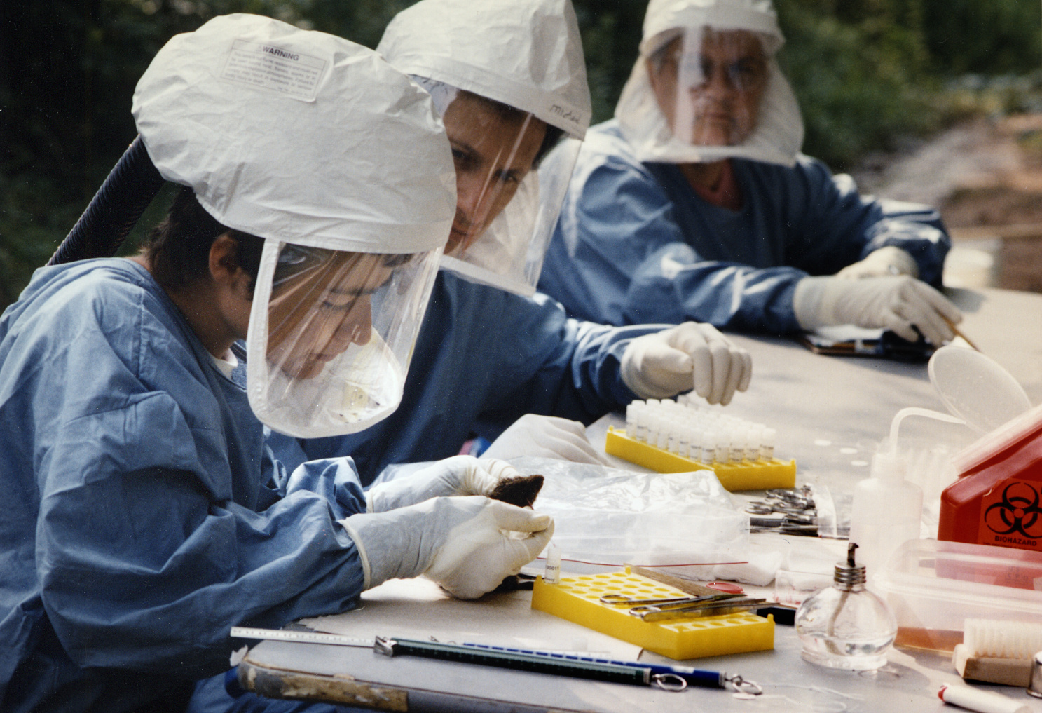
Image context: the cover is a real CDC Public Health Image Library photograph of three health officials inspecting suspected hantavirus specimens. Its catalogue record does not identify the outbreak, date, or place. That limit matters in an article about evidentiary chains: the photograph shows the situated work of field specimen processing, but it is not evidence that these individuals worked in Four Corners in 1993.[9]
May 14–28: two deaths become a surveillance problem
Malone and Tempest did not begin with a pathogen. They began with a pattern and a jurisdictional gap. The young woman's death at a clinic on the Navajo Nation had not entered New Mexico's state medical-investigator system; the young man's death in Gallup had. Once the household connection surfaced, the two men searched coroner records and called clinicians across the region. On May 17, the New Mexico Department of Health was alerted. A request then went to clinicians in Arizona, Colorado, New Mexico, and Utah for reports of similar cases. On May 28, state officials asked CDC for assistance.[3]
The first technical tool was not a virus assay. It was a case definition. Investigators agreed to examine people with otherwise unexplained bilateral lung infiltrates and low blood oxygen, as well as deaths involving unexplained noncardiogenic pulmonary edema. The initial CDC report made the threshold concrete: oxygen saturation below 90% while breathing room air, or a matching autopsy finding. By June 7, surveillance had found 24 provisional case-patients with illness onsets reaching back to December 1992; 12 had died. Those were suspected cases, not 24 laboratory confirmations—a distinction that kept the search broad without pretending uncertainty had vanished.[1]
The definition did more than count. It made old charts, radiographs, autopsies, and new admissions comparable across four states. It also prevented the investigation from being limited to a single household or a single ethnic label. Fourteen of the 24 early suspected patients were American Indian, but the list also included nine white patients and one Hispanic patient. Geography and exposure still needed explanation. An early demographic concentration was a clue about where surveillance had found illness, not evidence of an illness inherent to a people.[1]
May 29–June 4: the lungs contradict the known disease
CDC investigators arrived in Albuquerque less than a day after the request for help and joined staff from the University of New Mexico, Indian Health Service, state agencies, and other partners. On June 1, a 15-person CDC group began reviewing records, sending human tissue to Atlanta, interviewing families, and inspecting homes and workplaces. Plausible explanations included influenza, an environmental toxin, and an unknown pathogen. Plague, anthrax, and tularemia were among the alternatives tested against the accumulating evidence rather than dismissed by intuition.[3]
The patients' course was unusually compressed. The later clinical study described 18 confirmed infections, 14 of them fatal. Among 17 patients with records available, symptoms had lasted a mean 5.4 days before hospitalization. Fever and muscle pain could look ordinary at first; pulmonary edema, hypotension, a rising hematocrit, low platelets, and respiratory failure could follow with startling speed. The fatality pattern was not the result of a mild pneumonia being neglected for weeks. It was a new syndrome changing phase over days.[2]
On June 4, CDC's Special Pathogens Branch had results from an intentionally wide laboratory panel. Antibodies from all nine patients tested reacted with three known hantaviruses and with none of the other 22 viruses in the panel. That was a direction, not yet a complete identification. Known pathogenic hantaviruses were associated mainly with hemorrhagic fever and kidney injury in Eurasia. These patients had little of the expected renal picture; their dominant injury was a catastrophic leak across the lung's capillary barrier.[3]
This mismatch was scientifically productive. Investigators did not discard the antibody signal because the syndrome failed to resemble the textbook. They asked whether a previously unrecognized hantavirus could have a different organ target. The clinical evidence changed the category, while the category helped investigators read the clinical evidence.
June 10–16: a sequence in a person meets a sequence in a mouse
Serology established family resemblance. Molecular evidence tightened the link. On June 10, CDC scientists used reverse-transcription PCR to recover part of the suspected virus's RNA sequence from human material; viral antigen was also found in the endothelium lining pulmonary capillaries and other tissues. The genetic analysis published later that year placed the agent within the hantavirus family while distinguishing it from the viruses already known to science.[3][4]
At the same time, field teams trapped small mammals around case and comparison sites. On June 16, investigators reported the same viral sequence in deer mice, together with hantavirus antibodies in the animals. Human illness and animal reservoir were no longer connected only by the general fact that hantaviruses are rodent-borne. They were connected by matching molecular evidence from the outbreak landscape.[3]
The full reservoir study shows why the field operation was more than a dramatic mouse hunt. Investigators sampled 1,696 small mammals from 31 species. Deer mice were numerically dominant, had the highest antibody prevalence, and carried RT-PCR products indistinguishable from those recovered from patients. More than 96% of antibody-positive deer mice were also PCR-positive, consistent with persistent infection. The conclusion rested on convergence: abundance, serology, and sequence—not on finding one infected animal near one home.[5]
The household evidence added a crucial boundary. In a study of 17 case homes and matched comparison homes, median trap success was 17.3% at case sites, compared with 12.7% at nearby controls and 8.3% at more distant controls. Yet the proportion of antibody-positive deer mice did not differ significantly across those groups. What separated case households was chiefly more rodents, not a uniquely virulent pocket of mice. Exposure opportunity was the hinge.[6]
The wet winter explains pressure, not destiny
Why did recognition happen in spring 1993? The strongest ecological explanation begins with the wet 1992–1993 El Niño period. Greater precipitation increased vegetation and other food resources; rodent populations expanded, then mice encountered people and buildings more often as conditions shifted. A later Four Corners analysis reported a 20-fold rodent-population increase at central New Mexico's Sevilleta site over the previous year and found that the outbreak followed unusually high precipitation.[7]
That is an ecological mechanism, not a weather alibi. Rain did not itself transmit a virus, and a regional population boom did not make every home equally risky. The household study's density gradient, the field team's species testing, and the genetic match identify the links that the climate story alone cannot supply.[5][6][7] Nor was the virus necessarily born in 1993. University of New Mexico museum collections later found evidence of Sin Nombre virus in archived deer-mouse tissues gathered before the recognized outbreak. The event was an emergence into medical visibility, not proof of a sudden viral creation.[8]
The distinction protects against hindsight. Once a reservoir has been named, it is tempting to rewrite the investigation as an obvious line from wet weather to mice to disease. In May 1993, investigators still had to distinguish a toxin from influenza, a familial exposure from person-to-person spread, and a suggestive antibody reaction from a new pathogen. Ecology made the final account coherent; it did not make the intermediate tests unnecessary.
The name revealed a second outbreak cost
Scientific speed did not prevent social harm. As reports emphasized deaths among tribal members, Navajo and Hopi people were shunned in public places and excluded from regional events despite the absence of evidence that ordinary contact was spreading the illness. Later, when researchers proposed naming the virus for Muerto Canyon, Navajo objections made the problem unmistakable: a community already associated with fear would again be fixed to the pathogen in scientific language. The adopted name, Sin Nombre virus—Spanish for “virus without a name”—recorded that refusal.[3]
This is not a side story about public relations. Outbreak investigation depends on families permitting interviews and examinations, communities allowing field teams into homes, clinicians reporting across institutions, and local health authorities remaining credible. A technically correct message can still damage the cooperation on which its evidence rests if it turns a place or people into shorthand for contagion.
The response itself was broader than the popular image of outsiders arriving to solve a mystery. The first CDC report names the Navajo Nation Division of Health alongside the four state health departments, Indian Health Service, universities, medical examiners, and federal laboratories.[1] The discovery network worked because local clinical memory raised the alarm, tribal and regional partners made field investigation possible, and specialized laboratories could test what those partners collected. Giving one institution the whole victory would erase the system that produced the result.
What changed after the match
By July 27, 1993, 18 Four Corners patients had laboratory-confirmed infection and 14 had died. By October, surveillance had found confirmed cases well beyond the original region, proving that the risk could not sensibly be fenced inside a place-name. The clinical syndrome acquired a durable name—hantavirus pulmonary syndrome—and the new virus-reservoir pairing made prevention specific: reduce rodent entry, avoid stirring contaminated dust, and treat rodent evidence in enclosed spaces as an exposure problem rather than ordinary dirt.[2][3]
The deeper legacy is an outbreak architecture. A medical examiner notices that one death resembles another. A clinician calls beyond a single hospital. A provisional definition converts memory into surveillance. Pathology establishes what the lungs are doing. Serology finds a viral family; PCR distinguishes a new member. Field ecology finds the reservoir, while a comparison study identifies the exposure gradient. Community objection corrects a damaging name.
No single step “solved” Four Corners. The investigation succeeded when three evidence chains were made to meet—and when the mismatches between them were treated as questions rather than inconveniences. A lung disease that did not behave like known hantavirus illness, a virus found where human disease had not been recognized, and an ordinary mouse carrying an extraordinary clue all had to be held together. The decisive instrument was coordination: a death record, a human tissue sample, and a field specimen made to answer the same question.
Sources
- Centers for Disease Control and Prevention, “Outbreak of Acute Illness—Southwestern United States, 1993,” MMWR 42(22) (June 11, 1993) — the first surveillance report, provisional case definition, early case counts, and multiagency investigation.
- Jeffrey S. Duchin et al., “Hantavirus Pulmonary Syndrome: A Clinical Description of 17 Patients with a Newly Recognized Disease,” New England Journal of Medicine 330 (April 7, 1994) — PubMed record for the case definition, clinical tempo, laboratory pattern, and initial outcomes.
- Charles J. Van Hook, “Hantavirus Pulmonary Syndrome—The 25th Anniversary of the Four Corners Outbreak,” CDC Emerging Infectious Diseases 24(11) (2018) — detailed reconstruction of the alert, task force, laboratory sequence, field investigation, stigma, and naming.
- Stuart T. Nichol et al., “Genetic Identification of a Hantavirus Associated with an Outbreak of Acute Respiratory Illness,” Science 262 (1993) — PubMed record for the molecular identification and evolutionary placement of the outbreak virus.
- James E. Childs et al., “Serologic and Genetic Identification of Peromyscus maniculatus as the Primary Rodent Reservoir for a New Hantavirus in the Southwestern United States,” Journal of Infectious Diseases 169 (1994) — institutional record and abstract for the 1,696-animal reservoir study.
- James E. Childs et al., “A Household-Based, Case-Control Study of Environmental Factors Associated with Hantavirus Pulmonary Syndrome in the Southwestern United States,” American Journal of Tropical Medicine and Hygiene 52 (1995) — rodent-density comparisons at case and control homes.
- David M. Engelthaler et al., “Climatic and Environmental Patterns Associated with Hantavirus Pulmonary Syndrome, Four Corners Region, United States,” CDC Emerging Infectious Diseases 5(1) (1999) — precipitation, rodent abundance, seasonality, and the limits of the El Niño association.
- University of New Mexico Museum of Southwestern Biology, “Sin Nombre Hantavirus History” — institutional account of the 1993 field collaboration and evidence from archived deer-mouse tissues predating outbreak recognition.
- Centers for Disease Control and Prevention, Public Health Image Library, image 7271 — source record for the documentary photograph of CDC personnel inspecting suspected hantavirus specimens used as the article image.