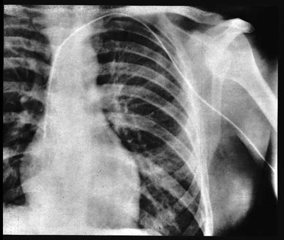
The thin pale line in Werner Forssmann's chest radiograph performs an unusually clean piece of persuasion. It rises from his left arm, disappears beneath the clavicle, bends down through the great veins, and ends at the shadow of the right heart. In November 1929, a reader did not have to accept the word of a 25-year-old surgical trainee that he had threaded a catheter from an arm vein into his own heart. The plate made the route visible.[1][2][8]
That image is so decisive that it has come to carry a much larger story. It is often treated as the birth certificate of modern cardiac catheterization: one forbidden experiment, one courageous doctor, one straight line to the catheter laboratory. But the radiograph settled only one of the necessary questions. It documented access to a living human right heart. It did not show that the procedure could be repeated safely across patients, that a sample taken there represented mixed venous blood, that pressure could be recorded accurately, or that the resulting numbers could answer a clinical question.
The more revealing microhistory therefore has several centers. Forssmann made a route undeniable in Eberswalde in 1929. Otto Klein in Prague soon used right-heart catheterization in patients to estimate cardiac output. At Bellevue, André Cournand and Dickinson Richards identified their sampling problem in 1936; after years of preparatory work, they and their collaborators began human cases in late 1940 and turned scattered precedents into a sustained system of sampling, pressure recording, calculation, and clinical interpretation.[4][11][12] The distinction does not shrink Forssmann's achievement. It explains what his X-ray proved—and what an X-ray, however memorable, could never prove alone.
A first does not appear from an empty past
Forssmann did not invent the idea of putting a tube into a heart. Nineteenth-century physiologists including Claude Bernard, Auguste Chauveau, and Étienne-Jules Marey had catheterized animal hearts to investigate pressure, temperature, and circulation. Human intravascular experiments also preceded him. Fritz Bleichröder and Ernst Unger passed long catheters through peripheral vessels in work reported in 1912; in one case they suspected that a catheter had reached the heart, but they did not confirm its position with a radiograph or a pressure trace.[2][6]
This is a useful boundary around the word “first.” Forssmann's claim was not that no catheter had ever traveled a long distance inside a person. His distinctive contribution was a documented human right-heart catheterization, deliberately undertaken for that purpose and published with visible evidence of the catheter's destination.[1][6] The evidence, not simply the distance traveled, made the episode durable.
Forssmann had just completed medical training when he arrived at the Auguste-Viktoria hospital in Eberswalde. He believed that venous access to the heart might permit more effective emergency drug delivery and might support contrast imaging of the cardiac chambers. His supervisor would not authorize an experiment on a patient. Forssmann made himself the subject instead.[1][2][6]
Later retellings—often following Forssmann's memoir—make the episode almost cinematic: a forbidden plan, sterile instruments obtained in secret, and a hurried trip to radiology with the catheter still in place.[10] Those details may describe what happened, but they reach us through retrospective narration rather than a source as immediate as the 1929 paper and plate.[1][10] The microhistory is stronger when the contemporary core and the later legend are kept in separate focus.
In 1929, the endpoint was an image
Forssmann exposed a vein near his left elbow, introduced a ureteral catheter, and advanced it approximately 65 centimeters. He then obtained the chest radiograph reproduced above. The Nobel biographical record and a later physiological history both describe the catheter as lying in the right auricle, the period term then used for the right atrium.[2][6] The point that mattered in 1929 was clear: a peripheral venous route could reach the right-heart region without the immediate catastrophe many physicians feared.
His brief paper did more than narrate a stunt. It connected the maneuver to possible intracardiac therapy and to contrast visualization. Forssmann continued that line of inquiry and in 1931 reported contrast imaging of the living right heart and pulmonary artery.[1][2][6] He had recognized that the catheter could be more than an object placed inside the body; it could carry a substance to a defined location and make otherwise hidden anatomy legible.
Still, survival of a single self-experiment is not a safety study. It cannot estimate an uncommon complication, reveal how a severely ill patient might respond, or separate the effects of the catheter from anxiety, movement, local anesthesia, and the surgical cutdown. Nor does willingness to accept a risk oneself establish that the same risk is justified for somebody else. Forssmann supplied a forceful feasibility demonstration. The work required after feasibility would be slower, more collective, and less photogenic.
The radiograph's great strength is also its narrative trap: one frame reduces the result to a visible yes or no. Everything that would make the result portable to other patients remained outside its border.
In Prague, the route yielded a number
Forssmann was not the only investigator to move quickly from possibility toward use. In 1929, Otto Klein at Prague's German University carried out diagnostic right-heart catheterizations in patients; his paper appeared in 1930. The American College of Cardiology's historical timeline records 11 procedures. Klein obtained what investigators then treated as mixed venous blood and applied the Fick principle to estimate cardiac output—the volume of blood pumped per minute.[11][12]
That work makes Klein more than a footnote between a daring route and its later refinement. Forssmann showed that a catheter could reach a conscious person's right heart and left radiographic proof. Klein demonstrated an early diagnostic use: the tube could furnish a sample for a physiological calculation. Yet his series did not seed a continuing research program comparable to the one later built at Bellevue, and histories organized around the 1956 Nobel laureates have often passed over it.[4][11]
Klein's place sharpens the distinction at the center of this microhistory. Scientific credit is not a ladder on which each rung belongs to one person. The sequence was not simply feat, then measurement. It was a documented feat, an early clinical calculation, and then the systematic consolidation that made results comparable across people, positions, instruments, and diseases.
At Bellevue, isolated uses became a program
Cournand and Richards approached the route from a different problem. Their Bellevue cardiopulmonary laboratory wanted a reliable sample of mixed venous blood so it could use the Fick principle to calculate how much blood the heart pumped. Samples from ordinary peripheral veins varied with the tissue draining into them. In 1936, they decided that a catheter advanced from a peripheral vein to the right atrium offered a direct way to sample the returning circulation.[4][5]
That sampling assumption was useful, but it was not final. Later studies showed that venous streams do not mix completely in the right atrium and can remain uneven in the right ventricle. In the absence of a vascular anomaly, blood from the pulmonary artery became the preferred sample for true mixed-venous oxygen content.[6] This correction belongs inside the success story: a productive measurement system must make its own assumptions testable.
The 1941 paper by Cournand and Hilmert Ranges begins by crediting Forssmann, then immediately turns to the objections that a usable method had to defeat. A foreign body might provoke thrombosis or thrombophlebitis. Clot could form inside the catheter. The procedure itself could disturb the subject enough to alter cardiac output—the very quantity under study. Their answer was not another spectacle but a specification: strict asepsis, defined local anesthesia, a flexible radiopaque catheter, controlled saline flow, observation, and measurements tied to position.[3]
The first report covered eight catheterizations in four people. Catheters remained in place for roughly 15 to 60 minutes without observed ill effects, and the team used right-atrial blood in a direct Fick calculation of cardiac output in one case.[4] Those denominators were still small. What changed was the shape of the claim. Forssmann had asked whether a catheter could arrive; Klein had shown that a sample could yield a cardiac-output estimate. Cournand and Ranges asked whether operators could make the catheter arrive consistently, leave it long enough to work, control foreseeable hazards, and recover a physiologically meaningful sample.
The method then accumulated layers. In 1941–1942, the group reported right-atrial pressure measurements and a series of 21 cardiac-output estimates. By the time a larger study was submitted in June 1944, the laboratory had performed about 260 human catheterizations. Curved tips improved positioning; double-lumen catheters allowed simultaneous sampling or pressure recording at two points; better manometers turned a mean pressure into a waveform.[4] No single refinement has the visual drama of the 1929 plate. Together they made the difference between access and an analytical system.
This multiplication of observations matters more than a simple growth in sample size. A pressure is useful only if the instrument is referenced correctly and the catheter tip is known to be in the intended chamber. A blood-gas value is useful only if the sample represents the circulation the calculation assumes. A repeated procedure is useful only if its complications, technical failures, and effects on the measurement are visible. The Bellevue work bound those dependencies together.
The years between cannot be cropped out
A heroic account commonly leaves Forssmann after the radiograph and brings him back for the Nobel ceremony. Biography cannot make that cut. A study based on material from his private archive found that in 1932 he joined the Nazi Party, the SA, and the Nazi Doctors' Association, before Hitler took power. Forssmann later served as a military medical officer. After the war, denazification proceedings barred him from medical practice for three years; the historians characterize his early political position as agreement with National Socialism followed by greater distance later in life.[7]
That record does not change where the catheter appears on the X-ray, and it does not erase the technical originality of documenting the route. It changes the moral frame around the familiar courage story. Voluntary self-experimentation is sometimes made to function as a certificate of character: the investigator risked himself, therefore the investigator was ethically admirable. Forssmann's fuller life shows why that inference fails. A person can accept bodily danger in one experiment and still align himself with a criminal political movement.
The opposite simplification fails too. A compromised biography does not make an empirical result disappear. The honest account holds both facts without asking either to launder the other. Forssmann's radiograph remains evidence. His political affiliations remain history. A health narrative worthy of the field has to preserve both.
The shared prize joined an experiment to a method
In 1956, the Nobel Prize in Physiology or Medicine went jointly to Forssmann, Cournand, and Richards for discoveries concerning heart catheterization and pathological changes in the circulation.[2][5] Its shared form matters: the award placed a singular self-experiment beside the sustained laboratory program that made the catheter scientifically productive. But it was not a complete map of the method's development; Klein was not among the laureates.[11]
Richards's Nobel lecture carefully described that conversion. By late 1940, the New York group could obtain consistent right-atrial blood-gas values, estimate cardiac output with the Fick principle, and keep the catheter in place long enough for extended study. Pressure recording and blood-volume methods followed. After a decade, the catheter was embedded in a set of methods that could investigate people with severe cardiopulmonary illness, not merely healthy volunteers.[5]
Richards also resisted another tempting origin myth. Surgery for congenital heart disease was already advancing independently; catheterization became a powerful aid because it could define abnormal anatomy and quantify the pressures and volumes of abnormal flow before and after an operation.[5] The tool did not single-handedly create modern heart surgery. It made certain questions answerable with new precision.
Nor should Forssmann's venous passage into the right heart be confused with every procedure now performed in a catheterization laboratory. Selective coronary angiography, percutaneous arterial access, balloon angioplasty, and stenting each required later investigators, devices, imaging systems, and evidence.[9] “He invented the modern cath lab” is a satisfying line only if most of the invention is removed.
The better legacy is more exact. Forssmann turned a proposed route into a documented event. Klein showed early that the catheter could serve diagnosis and calculation. Cournand, Richards, Ranges, and their colleagues turned scattered uses into a method whose risks and measurements could be inspected. Later clinicians turned that method into multiple diagnostic and therapeutic branches. The 1929 X-ray deserves its fame because it made one threshold impossible to deny. It becomes more meaningful, not less, when we stop asking it to contain everything that followed.
Sources
- Werner Forssmann, “Die Sondierung des rechten Herzens” (“Probing of the Right Heart”), Klinische Wochenschrift 8 (1929), 2085–2087 — the original report and bibliographic record of the self-catheterization.
- Nobel Prize Outreach, “Werner Forssmann — Biographical” — official biography covering his training, the 65-centimeter catheter passage, later work, and the 1956 award.
- André Cournand and Hilmert A. Ranges, “Catheterization of the Right Auricle in Man,” Proceedings of the Society for Experimental Biology and Medicine 46 (1941), 462–466 — primary report on the standardized technique, anticipated hazards, equipment, and sampling purpose.
- National Academy of Sciences, “André Frédéric Cournand,” Biographical Memoirs, vol. 67 (1995) — reconstruction of the Bellevue program, early case counts, measurement sequence, technical refinements, and the shift from access to an analytical system.
- Dickinson W. Richards, “The Contributions of Right Heart Catheterization to Physiology and Medicine,” Nobel lecture, December 11, 1956 — first-person account of the 1940s measurement program and the catheter's role and limits in cardiopulmonary medicine.
- Bobby D. Nossaman, Brittni A. Scruggs, Vaughn E. Nossaman, and Thomas J. Murthy, “History of Right Heart Catheterization: 100 Years of Experimentation and Methodology Development,” Cardiology in Review 18 (2010), 94–101 — open historical review of the animal and human precedents and the later measurement program.
- Lisa-Maria Packy, Matthis Krischel, and Dominik Gross, “Werner Forssmann — A Nobel Prize Winner and His Political Attitude before and after 1945,” Urologia Internationalis 96 (2016), 379–385 — archival study of Forssmann's Nazi affiliations, wartime correspondence, and denazification.
- Wikimedia Commons, “Werner Forssmann.jpg” — source page for the public-domain scan of the medical radiograph first published with Forssmann's 1929 paper.
- Martial G. Bourassa, “The history of cardiac catheterization,” Canadian Journal of Cardiology 21 (2005), 1011–1014 — overview separating right-heart catheterization from later coronary angiography and interventional milestones.
- Katharina Schroll-Bakes, “A rubber tube to the heart,” Siemens Healthineers MedMuseum (2022) — museum history that explicitly reconstructs the secret experiment from Forssmann's later autobiographical account and identifies the published radiograph.
- Pavel Gregor, Pavel Čech, and Petr Widimský, “History of cardiovascular research at the Charles University,” European Heart Journal Supplements 22 (2020), F1–F5 — open-access history identifying Otto Klein's 1929 diagnostic catheterization, mixed-venous sampling, and cardiac-output estimate.
- American College of Cardiology, “Our History” — institutional timeline recording Klein's 11 patient catheterizations and cardiac-output measurements in Prague.