Computed tomography now feels so ordinary that it is easy to misremember its arrival as a smooth technical upgrade from plain X-ray. The sharper history is more abrupt and more specific. CT crossed its real medical threshold when a machine built at EMI, installed at Atkinson Morley's Hospital in south London, made a previously hidden intracranial lesion appear as a readable slice of living tissue.[1][2][3]
That threshold matters because older brain imaging had a brutal bottleneck. The Science Museum's account of the EMI scanner states the problem plainly: before CT, X-rays could image the brain only after hazardous injections of air or special liquids.[1] Radiologists could infer, outline, or displace structures, but they could not yet inspect brain soft tissue directly with the kind of contrast modern clinicians take for granted. The first CT scanner mattered because it changed what the image itself was allowed to contain.
This reconstruction follows the hinge years from 1971 to 1974. The claim is narrower than "CT was invented." Hounsfield and Cormack later shared the 1979 Nobel Prize for the development of computer-assisted tomography.[5] But the first clinical turning point depended on something more crowded than invention alone: EMI engineering, James Ambrose's neuroradiology judgment, a prototype hospital installation, a first patient whose suspected frontal-lobe lesion could test the method, and a publication sequence that turned a local success into a new diagnostic regime.[2][3][4][5]
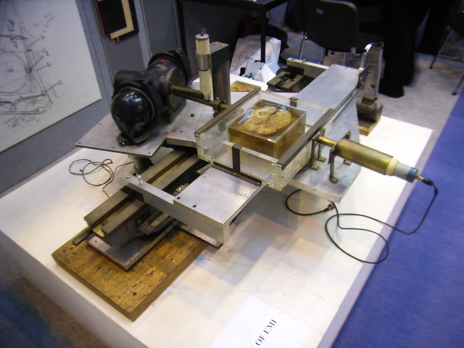
Image context: the cover uses a real photographic view of the first CT scanner prototype from Wikimedia Commons.[6] It belongs here because this story turns on physical machinery. The early scanner was slow, awkward, and narrow in scope, but it proved that brain pathology could be reconstructed from many X-ray measurements rather than guessed at from displacement signs.
Timeline anchors before CT felt inevitable
- 1 October 1971: the prototype EMI scanner was installed at Atkinson Morley's Hospital in Wimbledon.[2]
- 1971: the first patient, a 41-year-old woman with a possible frontal-lobe tumour, was scanned there; the data were sent back to EMI and the result returned after two days.[2]
- 12 October 1972: James Ambrose presented the new technique to the British Society of Neuroradiologists in Glasgow.[2]
- December 1973: the British Journal of Radiology published the three landmark CT papers, beginning with Hounsfield's "Part 1. Description of system."[2][3]
- January 1974: Atkinson Morley's team reported that the scanner had already been used on 650 patients.[2]
- 1975: JAMA described the EMI scanner as an exciting radiologic innovation capable of detecting minute alterations in intracranial absorption values.[4]
- 1977: the Science Museum notes that 1,130 CT machines had been installed worldwide.[1]
- 1979: the Nobel Prize in Physiology or Medicine was awarded jointly to Allan M. Cormack and Godfrey N. Hounsfield.[5]
1. Hounsfield's problem was not "pictures" in the abstract
The British Institute of Radiology's history page is useful because it keeps CT from sounding like a sudden miracle.[2] In the 1960s, Hounsfield was working at EMI on pattern-recognition problems and on the question of how a closed box with unknown contents might be examined from many directions.[2] That sounds abstract until the hospital collaborators enter. Frank Doyle supplied bone specimens, James Ambrose supplied brain specimens, and Louis Kreel supplied abdominal specimens, while EMI translated the concept into a workable prototype.[2]
That chain matters because the first CT scanner was not born as a universal imaging platform. It was narrowed into a solvable first task: the head.[1][2][3] The basic wager was that X-ray transmission readings taken through the skull at many angles could be reconstructed into absorption values for small internal units, then displayed as slices.[3] The early promise therefore sat less in speed than in a new way of organizing information. Conventional radiography projected structures onto each other. CT tried to separate them.
The original 1973 Hounsfield paper, even through its abstract alone, preserves this different ambition. It describes X-ray transmission readings taken through the head "at a multitude of angles," with absorption values calculated by computer and presented as pictures of slices of the cranium.[3] It adds the crucial comparative claim: the system was about 100 times more sensitive than conventional X-ray systems for displaying soft-tissue variations of nearly similar density.[3] That is the conceptual break. The scanner was not merely making prettier skull films. It was making soft-tissue difference itself newly legible.
2. Atkinson Morley turned the prototype into a hospital event
The event reconstruction becomes sharpest at Atkinson Morley's Hospital.[2] The prototype scanner was installed there on 1 October 1971. The scanning time was four minutes per slice, with a slice thickness a little over one centimetre. There was no computer attached to the machine itself. The data were recorded on magnetic tape and sent to EMI for analysis.[2]
Those details are what separate invention from clinical proof. The first patient was a 41-year-old woman with a suspected frontal-lobe tumour.[2] The data were acquired, the tapes went back to EMI, and the results returned after two days. According to the BIR account, the tumour was clearly shown, and Ambrose later said the result made him and Hounsfield jump up and down "like football players who had just scored a winning goal."[2] That reaction was not a laboratory celebration of elegant mathematics. It was the recognition that a lesion hidden inside a living brain had just appeared without the older violence of air studies or contrast displacement methods.[1][2]
This is why the first scanner's slowness should not be treated as an embarrassment. The machine took minutes per slice and days to reconstruct the image, yet it still changed radiology because the informational yield had shifted so dramatically.[2][4] A clinician could now ask a different question. Not merely: what has moved? But: what tissue occupies this slice, and how much does it attenuate compared with what should be there?
3. The 1973 papers made CT publishable, teachable, and comparable
Once the first scans worked, the next bottleneck was credibility beyond one hospital.[2][3][4] The December 1973 British Journal of Radiology issue mattered because it gave the method a formal triptych: Hounsfield on the system, Ambrose on clinical application, and Perry and Bridges on radiation dose.[2][3] That sequence is historically revealing. CT did not enter medicine as a single bragging image. It entered as hardware, clinical comparison, and dose accounting together.
Hounsfield's own article defined the system in operational terms: many-angle X-ray transmission measurements, computer-calculated absorption values, and sectional images of the cranium.[3] By 1975, the Mayo Clinic group writing in JAMA could describe computerized tomography as a recent radiologic innovation already useful for diagnosing intracranial lesions.[4] Their abstract states that a narrow moving beam of X-rays passed through the head, attenuation coefficients of small units of brain were plotted by computer, and the resulting displays showed axial slices capable of detecting minute alterations in intracranial absorption values.[4]
That language shows how quickly the field was learning a new image grammar. The interesting word is not simply "scanner." It is "coefficient," "attenuation," "slice," "minute alteration."[4] CT was teaching clinicians to read density differences numerically and anatomically at once. The brain was no longer just something outlined by indirect signs. It had become a layered object.
4. Scaling changed CT from a local triumph into a new default
The BIR history also shows why 1974 matters as much as 1971.[2] By January, the Atkinson Morley team had scanned 650 patients. The first year after installation had been devoted largely to evaluation; the second year moved the machine into full clinical use.[2] Resolution improved from an 80 x 80 matrix to 160 x 160, and the technique began to extend beyond the original proof-of-concept horizon.[2]
The first EMI scanner was still a head scanner, but the institutional consequences were already widening. The BIR page notes that following the success of the head scanner, the first body scanner was installed at Northwick Park Hospital, and the results transformed views of the body as well.[2] The Science Museum compresses the broader outcome into one blunt statistic: by 1977, there were 1,130 CT machines installed worldwide.[1]
That number matters because it shows how fast a hospital curiosity became infrastructure. A technology can be ingenious without becoming ordinary. CT became ordinary because the first clinical proof also generated a reproducible hospital workflow, a teachable publication record, and an expanding installed base.[1][2][3][4] By the time the Nobel Prize recognized Cormack and Hounsfield in 1979, the decisive professional argument had already been won inside radiology.[5]
What the first CT scanner actually changed
The early CT scanner did not make imaging instant, cheap, or universal.[1][2] It was slow, narrow, and tied to a specific hospital-engineering collaboration. But that is exactly why the Atkinson Morley episode deserves reconstruction. It shows the moment when the brain stopped being primarily a problem of projection and became a problem of sectional reading.
That is the lasting force of the first clinical CT scanner. Hounsfield's machine, Ambrose's radiologic judgment, and EMI's reconstruction pipeline together created a new medical object: the diagnosable slice.[2][3][4] Once that object existed, the rest of CT history could move quickly. Faster scanners, better matrices, body imaging, and eventually routine emergency and cancer staging all grew from that first proof that layered anatomy could be recovered from many-angle attenuation data inside a living patient.[1][2][5]
Sources
- Science Museum Group Collection, "EMI CT Brain Scanner" - official object page on the first clinically used EMI scanner at Atkinson Morley's Hospital, including 1971 installation context and the global installed-base figure by 1977.
- British Institute of Radiology, "Medical physics in the 1970s" - institutional history covering Hounsfield's EMI work, the Atkinson Morley installation on 1 October 1971, the first patient, four-minute slices, tape reconstruction, early patient volumes, and later technical scaling.
- G. N. Hounsfield, "Computerized transverse axial scanning (tomography): Part 1. Description of system" (British Journal of Radiology, 1973) - landmark paper abstract describing many-angle transmission readings, computer-calculated absorption values, sectional cranial images, and the roughly 100-fold sensitivity gain over conventional X-ray for similar-density soft tissue.
- H. L. Baker Jr. et al., "Computerized Tomography of the Head" (JAMA, 1975) - early clinical overview describing the EMI scanner's moving narrow beam, attenuation-coefficient plotting, axial-slice displays, and intracranial diagnostic usefulness.
- Nobel Prize Outreach, "The Nobel Prize in Physiology or Medicine 1979" - official Nobel summary for Allan M. Cormack and Godfrey N. Hounsfield.
- Wikimedia Commons, "File:RIMG0277.JPG" - source page for the article image showing the first CT scanner prototype developed at EMI.