The blood returns through a tube, looking much as it did when it left. Inside the dialyzer, however, its composition has changed. Dissolved wastes have crossed a membrane into a separate stream of dialysis fluid, and a controlled amount of water has left the circulation. Both changes happen in the same cartridge, which makes them easy to imagine as one operation.[1][2][3]
They depend on different driving forces. A concentration difference helps move dissolved substances; a pressure difference moves water. Understanding that distinction explains why clearing waste and removing fluid require separate attention during the same treatment.[2][3]
Keeping a difference along the membrane
A modern hollow-fiber dialyzer divides the flow into two compartments. Blood travels inside many fine tubes. Dialysis fluid, called dialysate, travels around their outer surfaces. The tube walls form the exchange membrane, allowing selected dissolved substances through while retaining blood cells.[1][2]
For a small waste molecule such as urea, removal depends on its concentration being higher in blood than in the fluid across the membrane. Molecules move in both directions, but the net movement is down that concentration difference. This is diffusion. A membrane permits the exchange; the difference supplies its direction.[2]
Fresh dialysate flows against the direction of blood. This countercurrent arrangement helps sustain a useful concentration difference along the cartridge. Blood nearing the outlet meets incoming dialysate, preserving the opportunity for further exchange even after some waste has already left.[2]
The fluid's composition matters as much as its movement. It contains a prescribed mixture of dissolved substances, and clinicians can adjust it when blood tests show too much or too little of particular minerals. The treatment therefore depends on what is present on both sides of the membrane.[1]
What moves the water
Removing excess water uses ultrafiltration. A pressure difference across the membrane drives fluid from the blood compartment toward the dialysate compartment. The care team sets a fluid-removal goal and a rate intended to approach the patient's target weight without removing fluid faster than the person can tolerate.[3]
Consider a simplified calculation: removing two liters over four hours averages 500 milliliters an hour; removing that same volume over two hours averages 1,000. The total is identical, but the hourly demand doubles. These are arithmetic examples, not treatment targets. They show why a volume on its own cannot describe the burden of a session.
The National Kidney Foundation connects overly rapid removal with low blood pressure and cramping. When a person cannot tolerate the required rate, a longer session or an additional treatment may be needed. Time changes how a fluid goal can be reached.[3]
A falling waste concentration and a falling body weight therefore answer different questions. Neither measurement, by itself, establishes that the other part of treatment has been adequate or well tolerated. That is the practical consequence of the two mechanisms.[1][3]
The distinction was there from the beginning
The early machines make the separation unusually visible. Willem Kolff's rotating-drum kidney of 1943 carried blood through cellophane tubing wrapped around a drum. The Medical Education Institute's dialysis museum explains that wastes could diffuse into the surrounding bath, while the device's low operating pressure prevented effective excess-fluid removal.[4]
The 1948 Kolff-Brigham redesign still used a membrane wound around a drum. By 1960, Fred Kiil's plate dialyzer arranged membrane sheets between grooved boards and used negative pressure on the dialysate outlet to remove excess fluid. Changes in shape and pressure control gave engineers different ways to accomplish the paired tasks.[4]
This history clarifies the word “filter.” A dialyzer can exchange dissolved substances without necessarily removing the volume of water a patient needs to lose. Improving one function does not automatically settle the other.
The water outside the blood circuit
The membrane also explains why dialysis depends on an unusually careful water supply. Separation of the two streams permits molecular exchange; it does not make the dialysate irrelevant to the patient's safety.
In its 2024 guidance, the CDC describes an average week's hemodialysis as involving exposure to 300–600 liters of water. That water supports treatment across the dialysis system; the figure is not the volume removed from the patient's body. CDC links adverse events to chemical or biological contamination in dialysis water and describes standards covering purification, storage, distribution and contaminant testing.[5]
The cartridge is consequently only the visible meeting point of a larger system. Safe exchange depends on the fluid arriving at its outer compartment as well as on the blood moving through its fibers.[5]
Hemodialysis replaces part of kidney function, and its prescription must account for the individual patient.[1] Following the two routes through the cartridge makes that prescription more intelligible: the concentration difference helps govern what crosses, while pressure and time help govern how much water leaves.
Sources
- National Institute of Diabetes and Digestive and Kidney Diseases, “Hemodialysis” — blood and dialysate flow, individualized solution composition and limits of kidney replacement.
- Fresenius Medical Care North America, 2008K Level I Training Manual, part 490114, revision E, section II, pages II-3–II-5 — membrane compartments, countercurrent flow and diffusion.
- National Kidney Foundation, “Ultrafiltration” — pressure-driven fluid removal, treatment duration and tolerance.
- Medical Education Institute, Home Dialysis Central, “Dialysis Machine Museum” — Kolff's 1943 machine, the 1948 Kolff-Brigham redesign and the 1960 Kiil dialyzer.
- Centers for Disease Control and Prevention, “Water Use in Dialysis,” March 26, 2024 — water exposure, contamination pathways and treatment-system standards.
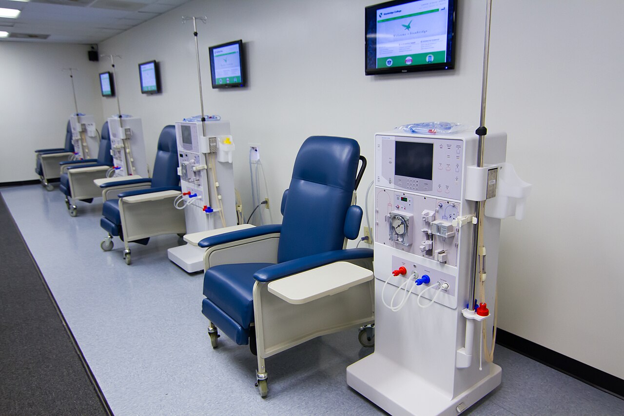
- Monirb, “Hemo 11-16-17.jpg,” December 4, 2012, Wikimedia Commons — Stanbridge College hemodialysis skills laboratory; original photograph and CC BY-SA 3.0 attribution.