The heavy apron at the dentist has always done two jobs. One is physical: a sheet of lead-equivalent material placed between a patient and possible radiation. The other is emotional: a ritual that says somebody is taking the invisible risk seriously. The myth begins when those two jobs are treated as the same thing. In modern dental radiography, the visible shield can feel safer while contributing little to the dose that matters and sometimes making the image harder to get right.[1][2][3]
The newer evidence story is not "radiation does not matter." It is almost the opposite. Radiation matters enough that the best protection is upstream: take an image only when it is justified, use prior images when possible, narrow the beam to the area being examined, position the patient correctly, choose lower-exposure techniques when they answer the clinical question, and avoid retakes.[1][5] The apron is familiar, but familiarity is not the same as benefit.
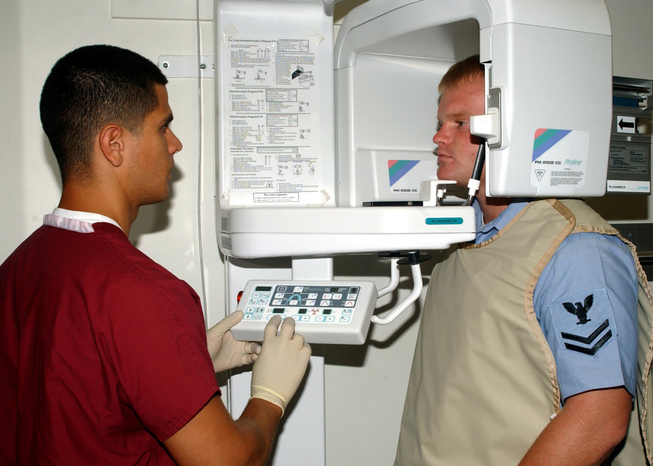
Image context: the cover uses a real 2005 U.S. Navy photograph of dental X-ray preparation.[6] It preserves the clinical room where this guidance has to be translated: a patient, a technician, equipment, and a trust problem. The article is about why the safety conversation has moved from a wearable shield to the whole imaging workflow.
Myth: if the apron blocks radiation, more apron must be safer
Lead is excellent at absorbing X-rays. That is why radiation workers still rely on distance, barriers, control booths, leaded windows, and protective garments when they face occupational exposure. Patient shielding during a dental image is a different problem. The patient is not standing next to the machine all day. The clinical task is to make one necessary diagnostic image with the smallest reasonable exposure and the lowest chance of repeating it.[1][3]
That distinction is why the American Dental Association's February 1, 2024 recommendations stopped treating abdominal aprons and thyroid collars as routine patient protection during dental X-rays. The ADA panel, supported by FDA medical physicists, concluded that the shields are not necessary for patients of any age or health status, including pregnancy, when modern dental imaging is performed appropriately.[1] The same release points to the stronger safety tools: digital imaging, beam restriction through rectangular collimation, good positioning, and using cone-beam CT only when lower-exposure options will not answer the clinical question.[1]
The apron myth survives because it makes intuitive geometry: if radiation travels outward, a barrier in front of the torso should help. But much of the residual exposure outside the target area in dental imaging is not a clean external beam heading toward the abdomen. It is scatter generated inside the patient after the beam has already interacted with tissue. A shield placed over the body cannot undo that internal scatter.[2][3]
Evidence: the dose question has been measured, not merely asserted
A useful example is the 2013 phantom study by Rottke and colleagues on panoramic radiography. The researchers used a full anthropomorphic body phantom with 110 thermoluminescent dosemeters placed at 55 sites, then compared scans with and without a standard adult lead apron on two panoramic devices.[4] The result was not a hidden protective effect waiting to be discovered. For both devices, the study found no statistically significant difference in absorbed doses with apron shielding versus without it.[4]
Those details matter because they make the argument less ideological. This was not a survey of patient preferences or a dentist's habit. It was a dose-measurement experiment. The numbers also show why old safety gestures can persist after their physical value has thinned. Panoramic radiography still feels like a machine moving around the whole head, so a torso shield feels sensible. The measured comparison says the torso shield is not where the meaningful protection is happening.[4]
The American Association of Physicists in Medicine makes the broader medical-imaging version of the same point. Its patient-shielding FAQ says routine gonadal and fetal shielding should not be the default and explains that shielding often provides negligible benefit compared with scatter generated within the body.[3] It also gives the practical reason clinicians worry about shields: if a shield covers anatomy needed for diagnosis, the image may need to be repeated.[3]
Myth: no shield means no safety culture
This is the emotionally important myth. Patients may reasonably wonder whether the office has become casual about radiation. The best answer is not to dismiss the worry. It is to name what replaced the apron as the center of safety.
The ADA's current oral-health topic page still frames radiographs as clinical decisions rather than routine commodities. It points clinicians and payers back to ADA/FDA patient-selection guidance, stresses clinical necessity, and says images should be correctly identified and diagnostically adequate rather than generated merely for administrative convenience.[5] That is a larger safety lever than a symbolic shield: do not take an image unless it answers a real question.
The 2024 ADA update sharpened the same logic. It did not say dental X-rays are casual. It said clinicians should order radiographs in moderation, use images acquired at prior dental exams when possible, restrict beam size, position patients properly, and follow applicable radiation-safety regulations.[1] In other words, the safety culture moved from the patient's lap to the prescription, equipment setup, and operator technique.
Evidence: shields can create their own failure mode
The argument against routine shielding is not only that aprons do little. It is also that they can sometimes make the exam worse. A thyroid collar or abdominal shield can block the primary X-ray beam or create artifacts. If the resulting image is nondiagnostic, the patient may need another exposure.[1][3] A safety item that raises the retake risk has to be judged by the whole episode, not by the comforting moment when it is placed on the patient.
Automatic exposure control is another reason the broader radiology world has moved away from routine patient shielding. AAPM explains that some modern imaging systems can change output when shielding enters the beam path, which can undermine the intended dose reduction.[3] Dental imaging does not reduce to one machine type, but the principle is general: modern safety depends on how the equipment, detector, beam, and patient position interact. A loose shield added late in the process can become a variable, not a guarantee.
The same is true for thyroid collars in dental imaging. The thyroid is close to the head and neck field, so older practice often treated a collar as obviously prudent. The newer AAOMR position, indexed in PubMed as a 2023 JADA recommendation, moved against routine gonadal, fetal, and thyroid shielding in dentomaxillofacial radiography.[2] That does not mean the thyroid is irrelevant. It means the better protection is beam restriction, selection criteria, image receptor improvements, and avoiding unnecessary images or retakes.[1][2]
The pregnancy and pediatric question
The hardest part of this transition is explaining it for children and pregnant patients. These are exactly the cases where patients and parents expect extra visible protection. The ADA's recommendation explicitly says the no-routine-shielding conclusion applies regardless of age or health status, including pregnancy.[1] AAPM's FAQ similarly says fetal and gonadal shielding should not be used by default, regardless of age, sex, or pregnancy status, while still allowing professional judgment for very anxious patients if image quality and dose are not compromised.[3]
That nuance matters. A clinician can explain that the apron is no longer the routine evidence-based safety tool and still treat anxiety as real. If a patient refuses a necessary image without shielding, the practical question becomes whether careful shielding can be used without covering anatomy or increasing dose. The point is not to win an argument about a garment. It is to get the needed diagnosis with the least overall risk.[3]
The child-specific concern should also be routed toward the strongest controls. Was the image clinically indicated? Could a previous image answer the question? Is the beam restricted? Is the patient positioned well enough to avoid a retake? Is CBCT being reserved for cases where two-dimensional imaging will not provide the necessary information?[1][5] These questions are less theatrical than placing a collar, but they are the safety questions with leverage.
Why the old ritual lasted
The lead apron lasted because it solved a communication problem. X-rays are invisible, cumulative-sounding, and historically associated with real harms. A visible barrier gave patients a role in their own protection. It also gave dental offices a simple script: we shield you because we are careful.
The science has now made the script harder. In 2013, the panoramic-radiography dose study found no significant apron effect.[4] In 2019, AAPM issued its position against routine gonadal and fetal shielding in diagnostic imaging.[3] In 2023, AAOMR published dentomaxillofacial shielding recommendations in JADA.[2] On February 1, 2024, the ADA announced updated dental radiography safety recommendations that no longer recommend routine lead abdominal aprons or thyroid collars.[1] The timeline is important because it shows a gradual migration, not a sudden whim.
The better replacement ritual is conversation plus technique. A dental team can say: we are not skipping protection; we are using the protection that works now. We take X-rays only when they change care. We use the smallest useful field. We position carefully. We avoid repeat images. We follow the law. We protect staff who face occupational exposure. And we will answer questions before taking the image.[1][3][5]
That is the myth-vs-evidence boundary. The lead apron was not foolish. It belonged to an older safety culture built around visible shielding. Modern dental radiography asks for a quieter discipline: justify the image, control the beam, trust measured dose evidence, and prevent retakes. The safest dental X-ray is not the one with the heaviest apron. It is the necessary image done correctly the first time.[1][4][5]
Sources
- American Dental Association, "ADA Releases Updated Recommendations to Enhance Radiography Safety in Dentistry" (February 1, 2024) - updated recommendation against routine lead abdominal aprons and thyroid collars, plus workflow safety measures.
- PubMed record for Benavides et al., "Patient shielding during dentomaxillofacial radiography: Recommendations from the American Academy of Oral and Maxillofacial Radiology," JADA, 2023 - AAOMR shielding recommendation record and DOI.
- American Association of Physicists in Medicine, "FAQs Patient Shielding" - patient-shielding rationale, negligible benefit, retake risk, pregnancy/pediatric boundary, and occupational-shielding distinction.
- D. Rottke et al., "Influence of lead apron shielding on absorbed doses from panoramic radiography," Dentomaxillofacial Radiology, 2013 - phantom dose study using 110 dosemeters at 55 sites.
- American Dental Association, "X-Rays/Radiographs" - current oral-health topic page summarizing patient-selection, clinical-necessity, diagnostic-quality, and payer-use guidance.
- Wikimedia Commons, "Dental X-Ray.jpg" - 2005 U.S. Navy photograph by Photographer's Mate 3rd Class Clarck Desire, used as the article image.