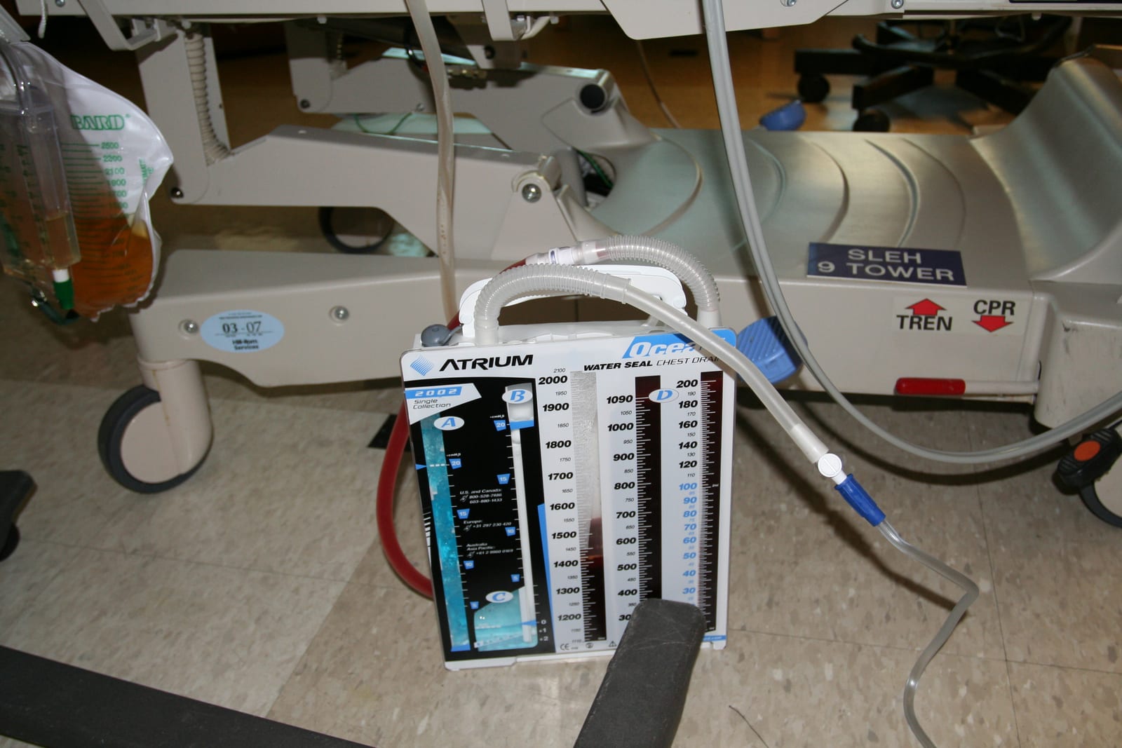
The least dramatic part of a chest drain is the part that makes it safe. A tube passes through the chest wall; a clear box sits below the bed; a few centimetres of water occupy one narrow chamber. The water does not pull a collapsed lung open. It does something more modest and more important: it gives air a route out without offering the room a route back in.
That distinction separates the mechanism from its common caricature. A chest drain is not simply a vacuum hose. Pressure gradients expel air, gravity assists fluid drainage, and separately prescribed suction may add force. The underwater seal makes the air path asymmetric. It is a door, not a pump.[2][3]
The problem is a broken pressure relationship
Each lung is covered by visceral pleura; the inside of the chest wall is lined by parietal pleura. Between them is normally a potential space containing only a thin film of fluid. The lung's elastic tissue tends to recoil inward while the chest wall tends to move outward. Their opposition helps keep pleural pressure below atmospheric pressure through much of normal breathing and couples the lung to the moving chest wall.[2][5]
Air, blood, pus, or other fluid in that space changes the relationship. In a pneumothorax, air separates the pleural surfaces and the lung recoils. In a pleural effusion or haemothorax, fluid occupies room the lung needs. A chest tube creates an exit, but an open tube alone would also create an entrance. During inspiration, subatmospheric pressure could draw room air toward the pleural space and preserve the very problem the tube was meant to relieve.
The drainage system therefore has to coordinate three jobs: carry air or fluid away, stop atmospheric air from returning through the tube, and allow the normal pressure relationship to re-form when the underlying lung can expand. Keeping the unit low guards separately against fluid reflux. The water seal is the low-tech air valve inside that system.[2][3][5]
A shallow column sets the direction
In the historical one-bottle arrangement, the tube from the patient ended beneath the surface of sterile water. Modern disposable units divide the work among connected chambers, but the seal keeps the same physical logic. Depending on the device, the submerged depth is around 2–3 centimetres or a manufacturer-marked fill line.[2][3]
When pressure on the patient side rises enough to overcome that shallow water column—as it may during expiration or a cough—air passes through the submerged opening and bubbles into the chamber. The chamber is vented, so that air can then escape to the atmosphere or into a separately connected suction system. “Closed drainage” does not mean a bottle sealed shut; it means the pleural space is protected from atmospheric air re-entry through the tube.
When pressure on the patient side falls, the water moves up the submerged tube instead of allowing room air to travel backward into the chest. The liquid column is therefore a hydrostatic threshold: outward air pressure can push through it, while attempted atmospheric air re-entry meets a fluid barrier.[2]
Fluid drainage exposes a second piece of design. In a single bottle, accumulating pleural fluid raises the water level and increases the depth the patient must overcome to expel more air. Two-bottle systems separate collection from the seal; contemporary boxes place those functions in adjacent compartments. The collection chamber can fill without silently changing the water-seal threshold.[2][5]
The unit stays below the drain insertion site for another reason. Elevation could permit collected liquid to run back toward the chest; a lower position supports gravity drainage and protects against that reflux. Upright position, adequate seal water, secure connections, and unobstructed tubing are not housekeeping details. They are conditions of the mechanism.[3]
The water is also a bedside sensor
A transparent seal reveals pressure changes. With spontaneous breathing, the water level may rise on inspiration and fall on expiration, a movement often called swinging or tidaling. Air leaving the pleural space may appear as bubbles. Long before a digital flow sensor, the chamber made an otherwise hidden pressure system visible.[2][3]
But visible does not mean self-interpreting. Persistent bubbling can represent a continuing leak from the lung, yet it can also come from a loose connection, a drain opening exposed outside the chest, or suction amplifying the flow. A sudden absence of swinging can mean the lung has re-expanded; it can also mean the tube is kinked, blocked, displaced, or disconnected from the pressure changes it should transmit.[3]
That ambiguity is why bubbling and swinging are observations, not verdicts. Clinicians interpret them alongside the patient's condition, the entire tubing circuit, drainage volume, imaging, and the reason the tube was inserted. A chest drain is an invasive hospital system; its chamber is not a prompt for a patient or visitor to clamp, reposition, refill, or troubleshoot it.
Suction belongs to a different chamber
Because the drain is often pictured beside wall suction, it is easy to credit the vacuum for everything. Yet the water-seal system can function without active suction: respiratory pressure gradients expel air, while gravity assists fluid drainage. When suction is required, traditional three-chamber systems regulate it in a separate chamber; newer dry or digital devices use a mechanical or electronic regulator. The wall source supplies airflow, while the drainage device limits how much negative pressure reaches the patient.[2][4]
This separation matters because more negative pressure is not automatically more treatment. The British Thoracic Society's 2023 guidance says robust evidence for routine thoracic suction is lacking outside particular settings, advises against applying it soon after insertion, and calls for prescribed, documented low-pressure suction when it is used. Excessive or premature suction can increase air flow through a leak or contribute to re-expansion pulmonary oedema rather than simply speeding recovery.[4]
The same causal logic explains one of the system's sharpest safety boundaries. If air is continuing to leak from the lung, the patent, unclamped drain is its escape path. Clamping that path can allow pressure to build into a tension pneumothorax. Current guidance therefore warns against clamping a bubbling chest tube except in specific circumstances under specialist pleural supervision.[3]
The important contrast is not “suction versus no treatment.” It is water seal versus suction as two different functions. The seal prevents atmospheric air from returning through the tube during ordinary breathing. Suction is an additional force, useful only when the clinical problem and the prescribed system call for it.
1871, 1875, 1967: the container changed, the rule survived
The familiar eponym gives Gotthard Bülau credit for closed underwater drainage, but the record is less tidy. In a February 1872 report, William Smoult Playfair described the continuous subaqueous drainage he had used for a child's empyema; historians date the intervention to 1871. Bülau independently used his own liquid-seal system in 1875 and published it in 1891, helping establish the method against an era of open drainage that accepted air entry as the price of draining infection.[1][5]
The next durable innovation was integration. By 1967, Deknatel had introduced a disposable unit that condensed the traditional collection, water-seal, and suction-control bottles into one bedside device. Digital systems now quantify air flow and pleural pressure, store trends, sound alarms, and provide portable suction. Some replace the visible water column with engineered valves.[2][5]
Technology changed the container, not the functional test. Any system still has to evacuate unwanted air or fluid, prevent reflux, manage pressure safely, and tell the care team enough to recognize failure. The 2006 bedside photograph looks old-fashioned beside a digital unit, but its placement and transparent chambers make those jobs unusually legible.[5][6]
A chest drain works because its components do not blur together. The tube provides a route. Pressure gradients expel air; gravity assists fluid drainage. The collection chamber preserves what has left for measurement. The water seal refuses atmospheric air's return journey. Suction, if needed, adds a controlled force rather than replacing the seal's logic.
The handful of water at the foot of the bed is therefore not incidental. It turns an open conduit into a directional system—and makes a hidden pressure relationship visible enough to watch.
Sources
- W. S. Playfair, “Paracentesis in Pleurisy and Empyema,” British Medical Journal 1(581), 17 February 1872 — the primary report of continuous subaqueous drainage through a tube leading to a water bottle below the bed.
- Charalambos Zisis et al., “Chest drainage systems in use,” Annals of Translational Medicine 3(3), 2015 — pleural-pressure mechanics, one-, two-, and three-bottle systems, the water seal, suction control, and the 1967 integrated unit.
- British Thoracic Society, “How to set up a chest drain bottle/underwater seal drain,” Online Appendix 8 to the 2023 Clinical Statement on Pleural Procedures — seal construction, positioning, observation, troubleshooting, and the clamping boundary.
- British Thoracic Society, “Suction and digital chest drain devices,” Online Appendix 10 to the 2023 Clinical Statement on Pleural Procedures — evidence limits, prescribed suction, pressure control, and digital-device boundaries.
- Claudio Sorino et al., “Chest Tubes and Pleural Drainage: History and Current Status in Pleural Disease Management,” Journal of Clinical Medicine 13(21), 2024 — historical development, functional requirements, chamber separation, and modern digital systems.
- Johntex, “Chest drain — bedside with fluids” (November 2006), Wikimedia Commons — source page for the clinical photograph used as the article image.