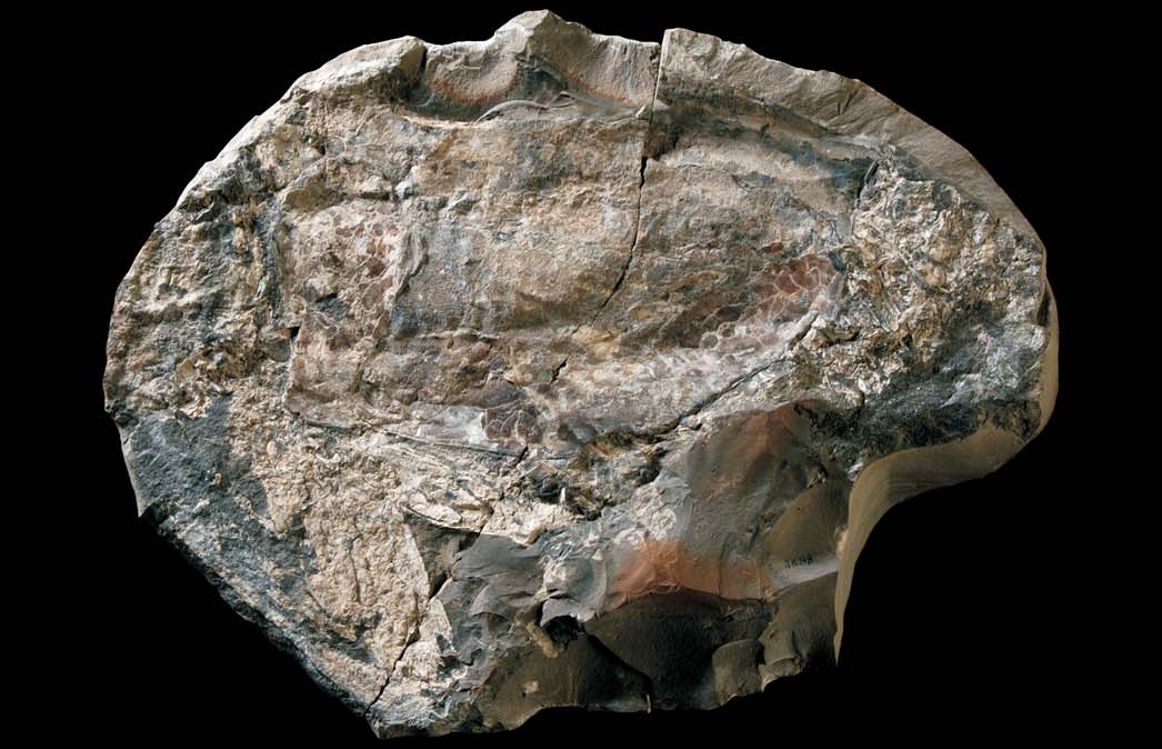
The eye commands attention before the animal makes sense. In specimen FSL 170759, a rounded mass crowds the front of Dollocaris ingens; jointed appendages reach out below it. Both the enormous eye and the grasping limbs are preserved anatomy.[1]
The animal came from La Voulte-sur-Rhône in southeastern France, a marine fossil deposit of the Callovian stage of the Middle Jurassic, approximately 165 million years old.[4] Its remains invite a deceptively simple question: what could it see? Answering that question changes how we imagine the water around it.
Looking inside the eye
Jean Vannier and colleagues’ 2016 study traced the eye beyond its outer surface. Each visual unit contained a lens, a crystalline cone and structures interpreted as receptor cells. Their arrangement identified an apposition eye, in which neighboring units sample different directions.[2]
The team estimated roughly 18,000 units per eye from preserved facets and assumptions about eye shape. That is an estimate, not a complete census. Together with grasping appendages and crustacean remains in the gut, the eyes supported an interpretation of Dollocaris as a visual predator.[2]
These clues answer different questions: capability, consumption and detection. Their agreement makes the hunting interpretation persuasive without preserving an actual pursuit.
Decay helped keep the details
Such fine anatomy raises another question: how did something so delicate become rock?
Clément Jauvion and colleagues investigated Dollocaris specimens preserved inside carbonate concretions. Their study, published online in 2019 and in a 2020 journal issue, identified phosphate minerals and iron sulfides replacing tissues, followed by a calcite cement that consolidated the structures. The chemistry varied with the kind of tissue being preserved.[3]
Their explanation gives decay an active role. Rapid microbial breakdown helped concentrate the ingredients for mineral precipitation inside the carcass. Under suitable local conditions, the process consuming the animal also helped replicate fragile structures before their shapes disappeared.[3]
Preservation was nevertheless selective. The specimens examined in that study lacked recognizable nervous and reproductive tissues. The authors cautioned that even exceptionally detailed fossils carry losses and distortions; missing anatomy cannot simply be read as anatomy the living animal never possessed.[3]
That changes the experience of looking at the photograph. The fossil's apparent solidity belongs to its later mineral history. To read the animal, we must also read the transformation that made it visible.
The seabed and the eye disagree
In their 2010 analysis, Sylvain Charbonnier and colleagues argued for a setting probably beyond 200 metres. Their case combined geological evidence with comparisons between the fossil community and living deep-water animals, including crustaceans, sponges, sea spiders and crinoids.[1]
Several kinds of organism appeared to point toward similar surroundings. Dollocaris was abundant in their sample, giving its habitat particular significance.[1]
The 2016 eye study introduced a difficulty. The optical anatomy suggested a need for moderate to strong illumination. Yet distortion and uncertain receptor dimensions prevented a reliable calculation of light sensitivity. The authors treated the conflict with a dark habitat as unresolved.[2]
A close reading should preserve that tension. The eye does not supply a depth gauge, and a resemblance to modern deep-sea communities does not give us a direct measurement of Jurassic light. The two arguments operate at different scales: one begins inside an animal, the other across a fossil assemblage. Neither becomes dispensable because the other is vivid.
A familiar eye in an unfamiliar body
Even the animal's family relationships have required another look. Dollocaris belongs to the extinct thylacocephalans, whose unusual body organization long complicated their placement among arthropods.[4]
In 2025, Thomas Laville and colleagues used synchrotron X-ray imaging to reveal previously unclear mouthparts and divisions of the body. They identified mandibles and two further pairs of mouthparts, and distinguished the anterior trunk carrying the grasping appendages from the posterior trunk. Their evolutionary analyses supported placement within Pancrustacea, close to malacostracans—the group containing crabs, shrimps and lobsters.[4]
That finding gives the animal a better-supported relationship without making it a Jurassic shrimp. Similarity in a conspicuous feature, such as a large compound eye, cannot do all the work of classification. Less theatrical structures around the mouth can be more revealing.
Return to the rounded eye and the reaching limbs. They let us recover something of an animal's encounter with its world: what it could detect, seize and consume. The brightness of that world remains a question. The fossil is compelling partly because the two kinds of knowledge have different limits.
Sources
- Charbonnier, Vannier, Hantzpergue and Gaillard, “Ecological significance of the arthropod fauna from the Jurassic (Callovian) La Voulte Lagerstätte,” Acta Palaeontologica Polonica (2010)—specimens, assemblage and environmental interpretation.
- Vannier and colleagues, “Exceptional preservation of eye structure in arthropod visual predators from the Middle Jurassic,” Nature Communications (2016)—eye anatomy, feeding evidence and optical uncertainties.
- Jauvion and colleagues, “Exceptional preservation requires fast biodegradation: thylacocephalan specimens from La Voulte-sur-Rhône (Callovian, Jurassic, France),” Palaeontology (2020; online 2019)—mineral replacement, decay and preservation bias.
- Laville, Forel, King and Charbonnier, “Synchrotron X-ray tomography sheds light on the phylogenetic affinities of the enigmatic thylacocephalans within Pancrustacea,” Proceedings of the Royal Society B (2025)—anatomical revision and evolutionary analysis; PubMed record.