Pulse oximetry is one of the most useful bedside technologies in modern medicine because it compresses a complicated physiologic state into a simple number. That simplicity is also the trap. A normal-looking SpO2 can persuade clinicians, patients, and family members that breathing is adequate when the device is only estimating oxygen saturation in pulsatile arterial blood.[1] It does not measure minute ventilation. It does not measure carbon dioxide clearance. It does not tell you whether the pulse signal is being weakened by vasoconstriction or shock. It does not rule out carbon monoxide poisoning. Read correctly, the pulse oximeter is a partial monitor with enormous value. Read lazily, it can turn delay into reassurance.[1][6]
The cleanest way to understand the device is to separate three bedside questions that often get collapsed into one:
- Is hemoglobin carrying oxygen?
- Is the patient ventilating well enough to clear carbon dioxide?
- Is the device getting a trustworthy arterial signal from this finger, on this patient, in this moment?
Pulse oximetry helps with the first question. It can mislead on the second. It can drift on the third.[1][2][4]
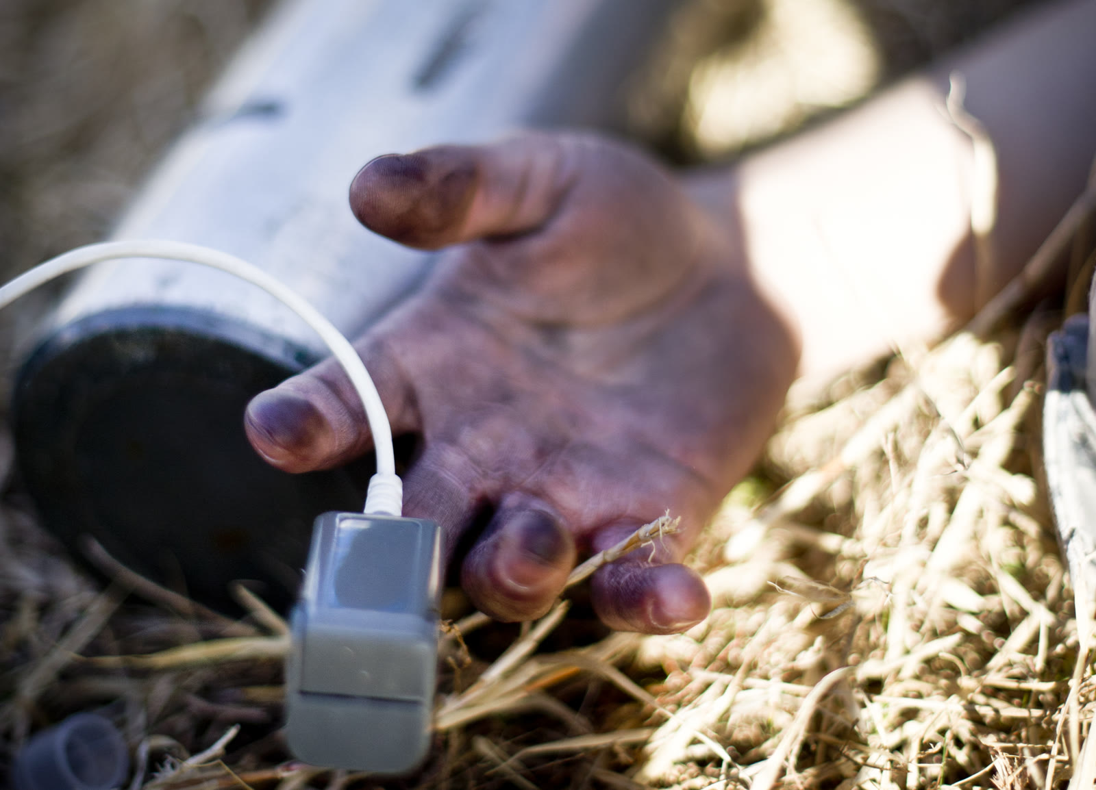
Image context: the cover shows a real fingertip pulse oximeter in use during a 2013 emergency-management drill at Yokota Air Base. It belongs here because pulse oximetry is a monitoring tool used inside clinical action, where the quality of the signal and the meaning of the number depend on the surrounding situation, not on the display alone.[7]
What the number actually is
The FDA's 2025 consumer update keeps the core definition simple: a pulse oximeter is usually clipped to a fingertip and uses light beams to estimate oxygen saturation and pulse rate.[1] That sentence matters because it places a boundary around the technology. SpO2 is an estimate, not a direct blood-gas measurement, and the estimate is only about oxygen saturation. The FDA also warns that readings must be considered alongside symptoms and that accuracy can be affected by poor circulation, skin pigmentation, skin temperature, skin thickness, tobacco use, and nail polish.[1]
That means the pulse oximeter is fundamentally an oxygenation monitor. It is useful when the clinical question is whether arterial hemoglobin is staying adequately saturated or whether that saturation is trending downward. It is much weaker when the clinical question is whether the patient is moving enough air. A patient can breathe too slowly, too shallowly, or with too much dead space and still preserve a reassuring saturation for longer than the room can afford.[1][2]
Why ventilation can fail before saturation does
The decisive misconception is treating oxygenation and ventilation as interchangeable. They are linked, but they are not the same physiologic job.
Ventilation is the movement of air in and out of the lungs, especially enough alveolar ventilation to clear carbon dioxide. Oxygenation is the loading of oxygen onto hemoglobin. A pulse oximeter tracks the second process. It does not directly report the first.[1][2] That distinction matters most during sedation, opioid toxicity, neuromuscular fatigue, and evolving respiratory failure, where carbon dioxide can rise before oxygen saturation visibly falls.
A 2004 Chest study made the masking effect of supplemental oxygen hard to ignore. During deliberate hypoventilation, SpO2 fell only in patients breathing room air; no decline occurred in those breathing inspired oxygen fractions of 0.25 or 0.30.[2] In the postoperative recovery setting, arterial desaturation below 90% was about fourfold more common in patients on room air than in patients receiving supplemental oxygen, 9.0% versus 2.3%.[2] The message was not that oxygen is harmful. It was that oxygen can hide ventilatory deterioration from the pulse oximeter by preserving saturation while carbon dioxide clearance worsens.
The same problem appears in procedural sedation. In a 2010 prospective colonoscopy study, capnography and pulse oximetry monitored 50 patients simultaneously.[3] Capnography found 29 apnea or hypoventilation episodes in 16 patients, with a mean event duration of 54.4 seconds. Pulse oximetry detected only 38% of those events, and when it did detect them, the mean delay was 38.6 seconds.[3] That is the clinical shape of the problem: the SpO2 display may stay calm precisely while the breathing problem is starting.
This is why a pulse oximeter should never be treated as a standalone breathing monitor in settings where hypoventilation is plausible. Respiratory rate, work of breathing, mental status, capnography when available, and blood-gas testing when needed remain separate tools because they answer separate questions.[2][3]
Why low perfusion turns measurement into a signal-quality problem
Even when oxygenation is the main question, pulse oximetry depends on a strong enough pulsatile signal reaching the sensor. The FDA's warning about poor circulation is not a minor footnote.[1] It names a core vulnerability. Cold hands, vasoconstriction, shock, low perfusion states, or motion can make the device's estimate drift upward or downward because the monitor is trying to extract an arterial pulse signal from a weakened or noisy optical field.
The 2024 prospective study by Gudelunas and colleagues adds more resolution to that warning.[4] In 146 healthy subjects, the investigators analyzed 9,763 matched pulse-oximeter and arterial-saturation readings across controlled hypoxemia. Low perfusion, darker skin pigmentation, and deeper hypoxemia all increased error. Under low-perfusion conditions, the combined frequency of missed hypoxemia diagnosis, defined as pulse-oximeter readings between 92% and 96% while arterial saturation was actually below 88%, was 1.1% for lightly pigmented skin, 8.2% for medium pigmentation, and 21.1% for dark pigmentation.[4]
Those numbers matter because they describe the exact failure mode that worries clinicians: a high-reading error, not a dramatic false alarm. The display does not collapse to zero. It looks acceptable while the arterial saturation is already in a range that should change management.[4]
Why hidden hypoxemia matters at outcome scale
Retrospective hospital data show that these discrepancies are not just a lab curiosity. In a 2022 multicenter cohort study, investigators analyzed 128,285 paired SpO2 and arterial-saturation measurements from 26,603 hospitalized adults.[5] Pulse oximetry overestimated arterial saturation by an average of 1.57%. Estimated occult hypoxemia, defined as arterial saturation below 88% despite pulse-oximeter saturation of 92% or higher, was 6.2% in Black patients versus 3.6% in White patients.[5] In ICU patients, occult hypoxemia was associated with increased odds of mortality, with an odds ratio of 1.36.[5]
The article's practical point is not to replace pulse oximetry with cynicism. It is to read the number as part of a risk model. When perfusion is poor, when skin-pigment-related error is plausible, or when the clinical exam looks worse than the screen, the number deserves verification rather than trust by default.[1][4][5]
Why carbon monoxide can make the device look reassuring
Carbon monoxide poisoning exposes the deepest boundary of conventional fingertip pulse oximetry because the question is no longer only oxygen saturation. It is abnormal hemoglobin chemistry.
The CDC's 2024 clinical guidance states this plainly: the key confirmatory test is carboxyhemoglobin measurement, usually by laboratory CO-oximeter, and the conventional two-wavelength pulse oximeter is not accurate when COHgb is present.[6] A fingertip pulse CO-oximeter can measure COHgb, but the standard bedside device most people mean by "pulse ox" cannot.[6]
That is why a patient with carbon monoxide exposure can look falsely reassured on a conventional device. The screen may show an acceptable oxygen-saturation number while the real toxic problem is that hemoglobin binding sites are occupied by carbon monoxide rather than oxygen. The CDC therefore anchors management around exposure history, symptoms, COHgb testing, and treatment with 100% oxygen, not around ordinary fingertip SpO2 alone.[6]
How to read a pulse oximeter correctly
The device becomes much more useful once its job is narrowed.
- Use it as an estimate of oxygen saturation trend, not as proof that breathing is adequate.[1][2]
- In suspected hypoventilation, treat normal SpO2 as insufficient reassurance, especially if supplemental oxygen is running.[2][3]
- In low-perfusion states, cold extremities, shock, or discordant clinical exams, question the signal quality before trusting the displayed value.[1][4]
- In patients at risk for hidden hypoxemia, especially when treatment thresholds are tight, remember that small overestimates can matter.[4][5]
- In suspected carbon monoxide poisoning, do not use a conventional pulse oximeter to rule the diagnosis out; get COHgb testing or pulse CO-oximetry if available.[6]
The pulse oximeter remains a strong monitor because oxygenation is important and continuous bedside trending is valuable. The correction is simply that one monitor cannot stand in for the whole respiratory exam. The number tells part of the story. The rest still belongs to physiology, context, and verification when the screen looks calmer than the patient does.[1][2][4][5][6]
Sources
- U.S. Food and Drug Administration, "Pulse Oximeter Basics" (content current as of March 26, 2025) - device definition, signal limitations, symptom context, and accuracy factors including poor circulation and skin pigmentation.
- Fu ES, Downs JB, Schweiger JW, Miguel RV, Smith RA, "Supplemental oxygen impairs detection of hypoventilation by pulse oximetry" (Chest, 2004) - deliberate hypoventilation and PACU data showing how supplemental oxygen can mask ventilatory deterioration on SpO2.
- Cacho G, Pérez-Calle JL, Barbado A, et al., "Capnography is superior to pulse oximetry for the detection of respiratory depression during colonoscopy" (Revista Española de Enfermedades Digestivas, 2010) - prospective sedation study comparing delayed SpO2 detection with real-time capnography.
- Gudelunas MK, Lipnick M, Hendrickson C, et al., "Low Perfusion and Missed Diagnosis of Hypoxemia by Pulse Oximetry in Darkly Pigmented Skin: A Prospective Study" (Anesthesia & Analgesia, 2024) - prospective evidence on the interaction of low perfusion, pigmentation, and high-reading error.
- Wong AI, Charpignon M, Kim H, et al., "Disparities in Hypoxemia Detection by Pulse Oximetry Across Self-Identified Racial Groups and Associations With Clinical Outcomes" (Critical Care Medicine, 2022) - multicenter cohort study linking occult hypoxemia with racial disparity and worse outcomes.
- U.S. Centers for Disease Control and Prevention, "Clinical Guidance for Carbon Monoxide Poisoning Following Disasters and Severe Weather" (July 8, 2024) - COHgb confirmation, 100% oxygen treatment, and the inaccuracy of conventional two-wavelength pulse oximeters when COHgb is present.
- Wikimedia Commons, "File:Yokota pulse oximeter.jpg" - source page for the archival photograph used as the article image.