MRI is often remembered as if it appeared fully formed: a patient slides into a cylinder, soft tissues bloom into view, and modern imaging simply begins. The better reconstruction is more mechanical and more interesting. MRI crossed its real clinical threshold only when three separate problems stopped living on separate timelines. First, someone had to show that magnetic resonance could encode location rather than only chemistry. Then someone had to make the method fast enough to image living anatomy instead of static phantoms and heroic long exposures. After that, hospitals still needed proof that the resulting contrast would change diagnosis in places where x-ray and CT remained weaker, especially in soft tissue.[1][2][3][4][5]
That sequence is why 1973-1983 is the right frame.[1][2][3][4][5] Paul Lauterbur's 1973 paper supplied the first decisive location trick. Peter Mansfield and Andrew Maudsley's fast-scan work in the mid-1970s supplied speed and anatomical legibility in vivo. Early-1980s British clinical work then showed that MRI was no longer a beautiful physics demonstration waiting for a medical use case. It had become a diagnostic instrument with a clear lane of superiority: non-invasive, non-ionizing, soft-tissue-heavy imaging.[2][3][4][5][6]
That is why MRI still reads sharply in 2026. Its success was not a single invention with a single date. It was a staged convergence between physics, engineering, and clinical demand.[1][2][3][5][6]
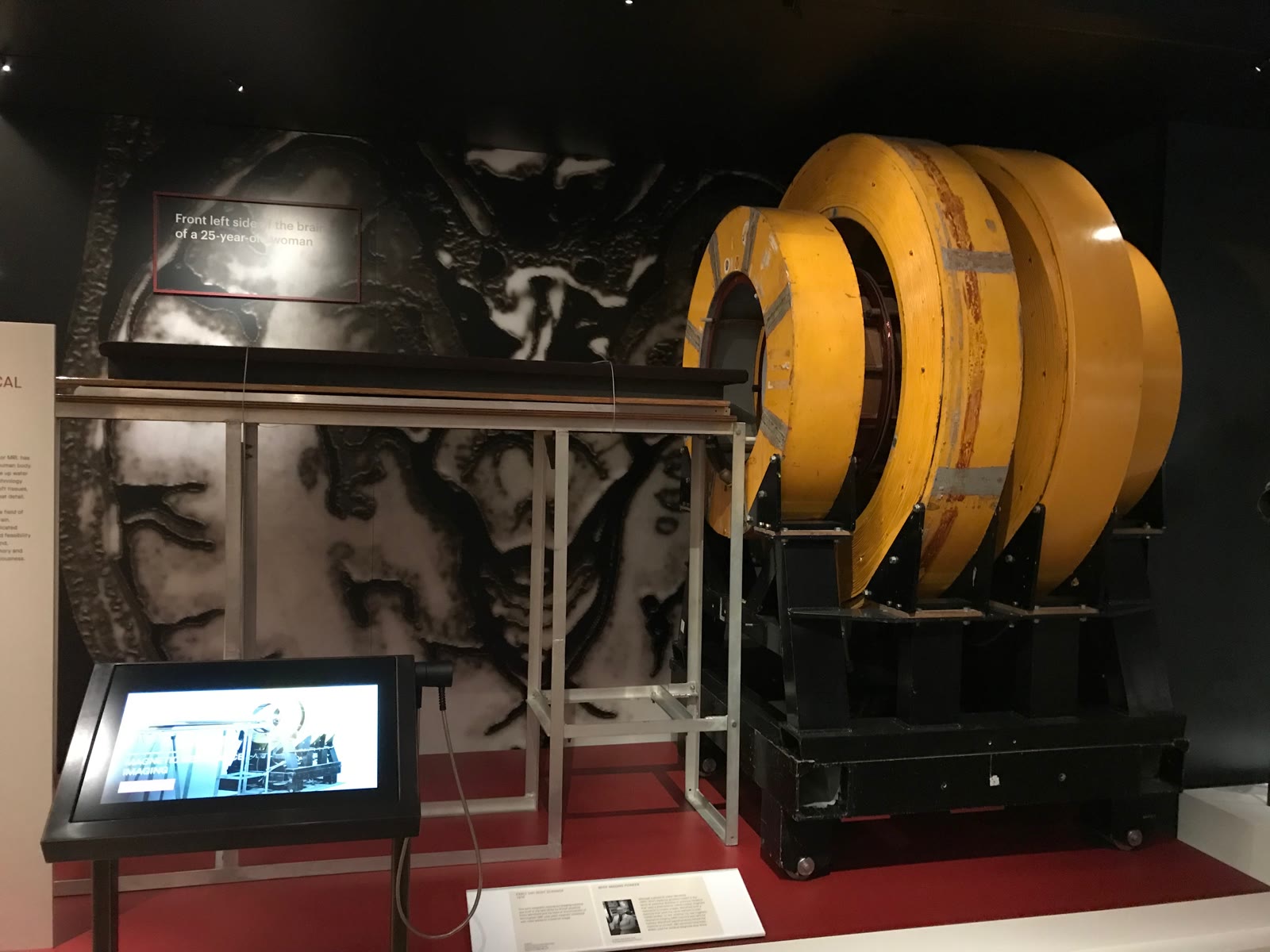
Image context: the cover uses a real photograph of the first MRI machine now displayed at London's Science Museum.[7] It fits this article because the threshold being reconstructed here was infrastructural. MRI became medicine only after an unwieldy experimental device became a room-sized clinical habit.
Timeline anchors before the machine looks inevitable
- 1973: Paul Lauterbur published his Nature proof of image formation by induced local interactions, showing that magnetic resonance signals could be spatially encoded through gradients and reconstructed into images.[1]
- 1977: Peter Mansfield and Andrew Maudsley published fast-scan proton imaging of a human finger, reporting considerable anatomical detail in vivo, especially in soft tissues.[2]
- 1980: the British Institute of Radiology's historical summary places the first clinical MRI produced in the UK in Nottingham and Aberdeen that year.[5]
- 1981: Roger Ordidge, Peter Mansfield, and Richard Coupland published the first biomedical images produced by high-speed echo-planar imaging, with clear anatomical detail in human and animal studies performed in vivo.[3][5]
- 1983: D. M. Kean and colleagues published clinical MRI examples of normal knee anatomy and pathology, an early sign that MRI had become a usable soft-tissue diagnostic tool rather than a laboratory curiosity.[4][5]
- 2003: the Nobel Prize in Physiology or Medicine recognized Lauterbur and Mansfield for the discoveries that made modern MRI possible.[6]
Those dates keep the story disciplined. MRI did not become clinically important the moment anyone imagined a scanner. It became clinically important when location, speed, and diagnostic contrast finally arrived in the same machine family.
1. Lauterbur solved the first bottleneck by making location visible
Magnetic resonance already had scientific value before MRI. It could tell chemists and physicists about molecular environments, relaxation properties, and signal behavior. That was powerful, though it was not yet anatomy.[1][6] Hospitals did not need one more elegant way to describe bulk material properties. They needed a way to say where a signal came from inside the body.
Lauterbur's 1973 breakthrough mattered because it changed that grammar.[1] The Nature paper described image formation by using magnetic-field gradients so resonance information could be linked to position, then reconstructed from multiple projections.[1] That may sound technical in summary, though the clinical implication was straightforward. Once resonance could be spatially encoded, the method no longer had to remain a tube-average or sample-average measurement. It could begin to behave like imaging.
This was the first threshold and also the narrowest one. Lauterbur did not deliver a finished hospital workflow in 1973.[1][6] He delivered proof that magnetic resonance could, in principle, leave the test tube and enter geometry. Without that step, everything later in MRI would have remained impossible or at least incoherent. A hospital scanner without location is not a scanner. It is spectroscopy pointing at a body.
2. Mansfield's group solved the second bottleneck by making living anatomy practical
Location alone was not enough. Early MRI could still have died as a slow, elegant method that worked beautifully on phantoms and badly on breathing, moving patients. Mansfield's contribution sits here.[2][3][6]
The 1977 British Journal of Radiology paper by Mansfield and Maudsley is historically valuable because it already sounds closer to medicine than to laboratory spectacle.[2] The abstract describes fast-scan proton imaging of a human finger in vivo and says the images revealed considerable anatomical detail, particularly in soft tissue regions.[2] That wording is the hinge. Once living anatomy could be shown with recognizable soft-tissue structure, MRI's future stopped depending only on imagination.
Speed then became the real engineering battleground. In 1981, Ordidge, Mansfield, and Coupland published the first biomedical images produced by high-speed echo-planar imaging, again emphasizing clear anatomical detail in vivo and even discussing the possibility of imaging moving objects and flow.[3] This is the harder second threshold. A technique that takes too long, blurs too easily, or cannot follow physiology will struggle to become everyday clinical infrastructure even if its underlying contrast is attractive.
That is why MRI history should not be compressed into one discovery date. Lauterbur made position legible.[1] Mansfield's group kept pushing the method toward acquisition speeds and image formation strategies that living bodies could tolerate.[2][3][6] Clinical imaging needed both achievements. Hospitals cannot diagnose from a principle alone. They need a machine that can finish the exam before the patient, the organ, or the workflow defeats it.
3. The early 1980s turned promise into a diagnostic lane
The best sign that MRI had crossed into medicine is not simply that the images existed. It is that clinicians started publishing anatomically and diagnostically targeted papers.[4][5] The British Institute of Radiology's 1980s history makes this shift easy to see in sequence: first clinical MRI in Nottingham and Aberdeen in 1980, high-speed biomedical images in 1981, then a clinical knee paper from Nottingham in 1983.[5]
The 1983 knee paper is especially important because it shows MRI finding a domain where its advantages were immediately legible.[4][5] Knee diagnosis depends heavily on soft-tissue differentiation: menisci, ligaments, cartilage, marrow-adjacent change, and pathology that plain x-rays do not show directly and CT of that era did not render as naturally.[4][5] Once MRI could publish normal anatomy and pathology in such a region, it had stopped being only a physics triumph. It had acquired a service line.
That service line also explains why MRI did not replace every other modality all at once. NIBIB's current overview states the contrast plainly even now: MRI is particularly well suited for non-bony soft tissues and differs from CT because it does not use ionizing radiation.[6] Brain, spinal cord, nerves, muscles, ligaments, and tendons are seen more clearly with MRI than with regular x-rays and often more clearly than with CT for specific questions.[6] This was the durable clinical bargain. MRI would earn its place where soft-tissue contrast justified cost, time, and complexity.
The event reconstruction therefore has a concrete endpoint. By 1983, MRI had enough of a workflow, enough speed, and enough diagnostic clarity that specialists could begin to treat it as a practical modality rather than a speculative one.[3][4][5]
4. MRI changed medicine by reordering what could be seen safely and repeatedly
This point matters because it keeps the article from turning into inventor worship. MRI won for a structural reason.[1][2][3][4][5][6] It let physicians inspect soft tissue in detail without cutting the body and without the ionizing radiation burden that came with x-ray-based imaging.[6] That combination created new habits in neurology, musculoskeletal imaging, oncology staging, and later functional and vascular applications.
The benefit should still be stated with boundaries. MRI was expensive. It was technically demanding. Motion remained a serious obstacle. Early systems were primitive by current standards.[5][6] Even now, MRI requires stillness, a powerful magnet, careful implant screening, and more time than many x-ray or CT studies.[6] The machine's history is therefore not a simple story of one superior image replacing inferior ones. It is a story of one modality finding the places where its specific contrast logic was worth the operational burden.
That is exactly what good medical technologies do when they last. They do not win by being abstractly modern. They win by solving a narrow, repeated problem better than the alternatives. MRI's repeated problem was soft tissue.
Why the 1973-1983 threshold still matters in 2026
The decade reconstructed here still feels current because hospitals continue to live inside the settlement it created.[5][6] When clinicians order MRI for the brain, spine, knee, shoulder, pelvis, or liver, they are still cashing in on the same three-part convergence: gradients that localize signal, acquisition strategies fast enough for living anatomy, and contrast properties rich enough to justify the scan.
That is the cleanest way to remember MRI. It did not arrive when one paper proved a possibility. It arrived when possibility became workflow. Lauterbur gave the method coordinates.[1] Mansfield's work made those coordinates usable in living tissue.[2][3] Early clinical papers then showed that medicine had somewhere real to put the machine.[4][5] By the time the Nobel committee honored MRI in 2003, the decisive threshold had already been crossed two decades earlier.[6]
Sources
- Paul C. Lauterbur, "Image Formation by Induced Local Interactions: Examples Employing Nuclear Magnetic Resonance" (Nature, 1973) - the foundational spatial-encoding paper that turned magnetic resonance from a bulk signal into an imaging method with reconstructable position information.
- P. Mansfield and A. A. Maudsley, "Medical imaging by NMR" (British Journal of Radiology, 1977) - fast-scan proton imaging of a human finger in vivo, with notable soft-tissue detail.
- R. J. Ordidge, P. Mansfield, and R. E. Coupland, "Rapid biomedical imaging by NMR" (British Journal of Radiology, 1981) - first biomedical images from high-speed echo-planar imaging, emphasizing in-vivo anatomical detail and the possibility of imaging motion and flow.
- D. M. Kean, B. S. Worthington, B. J. Preston, E. J. Roebuck, H. McKim-Thomas, R. C. Hawkes, G. N. Holland, and W. S. Moore, "Nuclear magnetic resonance imaging of the knee: examples of normal anatomy and pathology" (British Journal of Radiology, 1983) - an early clinical paper showing MRI's emerging diagnostic value in soft-tissue musculoskeletal imaging.
- British Institute of Radiology, "Diagnostic imaging in the 1980s" - historical overview used here for the early UK clinical timeline, including Nottingham and Aberdeen in 1980 and the rapid move into knee imaging by 1983.
- Nobel Prize Outreach, "The Nobel Prize in Physiology or Medicine 2003" - official summary of why Paul Lauterbur and Peter Mansfield were recognized, plus the broad diagnostic importance of MRI.
- Wikimedia Commons, "File:The first MRI machine at the Science Museum.jpg" - source page for the documentary photograph used as the article image.