A small probe rests between the ribs on the right side of the chest. The person being examined lies back, with the right arm raised to open the space. There is gel, light pressure and a succession of taps. No needle enters the liver. Yet the examination can help answer a question once closely tied to a tissue sample: how much scarring has accumulated inside?[1][6]
The answer begins with a measurement of stiffness. That distinction matters because a liver can become stiffer for more than one reason. To understand a FibroScan result, follow what travels from the probe into the tissue—and what has to be inferred on the way back.
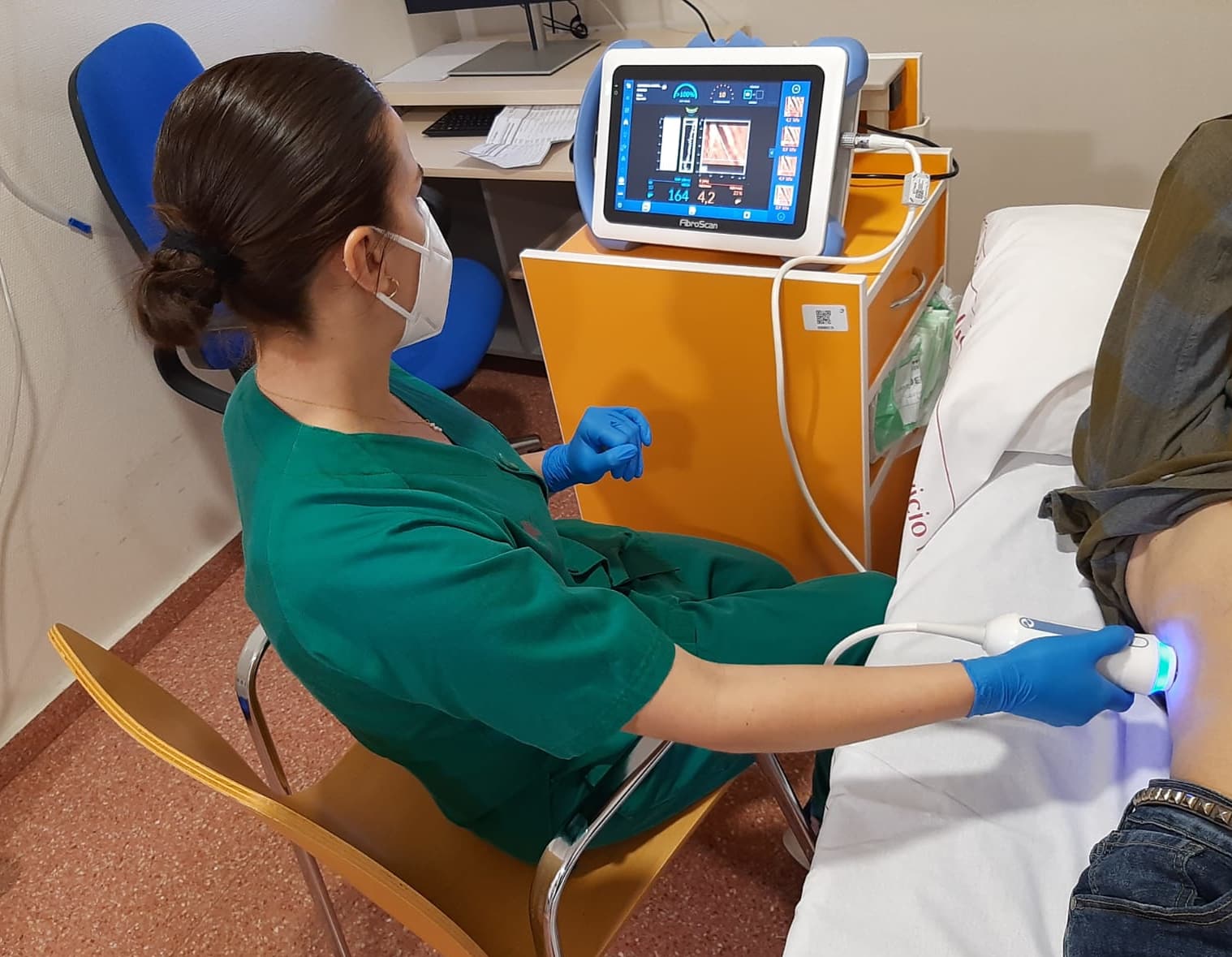
The tap and the listening instrument
Transient elastography combines two jobs. A mechanical vibration sends a shear wave through the liver; ultrasound tracks its passage. The wave travels faster through stiffer tissue. Software converts that speed into an estimate expressed in kilopascals, or kPa. The instrument therefore measures a physical property that can provide evidence about fibrosis, the buildup of scar tissue.[1][3]
In December 2003, Laurent Sandrin and colleagues reported an evaluation involving 106 patients with chronic hepatitis C. Their device paired a low-frequency vibration with ultrasound and found a relationship between measured stiffness and fibrosis grade. The important achievement was making a hidden tissue property accessible through the skin. The study established a useful association in a defined patient population; it did not establish that every stiff liver has the same cause.[3]
The distinction is easy to lose because the examination ends with a tidy number. A fibrosis stage, however, describes tissue damage. A stiffness reading describes how tissue responds to a wave. Moving from one to the other requires evidence about the patient as well as the machine.
Repetition makes a reading more dependable
One tap is insufficient. The 2018 CDC examination manual for the National Health and Nutrition Examination Survey specifies at least 10 valid measurements, with the reported stiffness represented by their median. It also checks how widely the readings are spread: its protocol uses an interquartile-range-to-median ratio below 30% when completing the examination.[1]
These checks address a practical problem. A plausible-looking central value can hide inconsistent measurements. Repeating the acquisition helps establish that the instrument is obtaining a dependable signal from the examination site.
But consistency cannot explain the signal's cause. Repeated measurements can agree closely while reflecting a temporary physiological change. Precision answers whether the readings hang together; it cannot, by itself, tell the clinician why the liver is stiff.
When drainage changed the number
A study published in November 2008 made that limitation unusually tangible. Gunda Millonig and colleagues examined patients with obstruction to bile drainage outside the liver. They measured stiffness before an endoscopic procedure and again 3–12 days after successful drainage. The reading fell in 13 of 15 patients.[4]
In three patients, the initial readings had reached levels suggesting advanced cirrhosis. Their mean stiffness fell from 15.2 to 7.1 kPa after drainage.[4]
Those changes showed that obstruction itself could contribute to stiffness. The small study did not establish a universal correction formula, or show that a large burden of scar tissue had disappeared within days. It demonstrated why the circumstances of a measurement belong beside the number.[4]
A meal can enter the measurement
The same reasoning reaches into an ordinary appointment instruction: fasting. In April 2017, Maria Kjærgaard and colleagues reported a study of 60 patients, most with alcohol-related liver disease. Participants received liquid meals containing 625 or 1,250 calories on separate study days. Researchers followed stiffness over the next three hours using both transient elastography and another ultrasound-based technique.[5]
Stiffness increased after both meals. Some readings crossed thresholds that would have assigned patients a more advanced fibrosis category. In some participants, the increase persisted through the final measurement. The liver's response to eating had entered a test being used to assess chronic damage.[5]
The study's selected population and liquid meals limit how precisely its results describe any one person's breakfast. They nevertheless explain why preparation is part of measurement quality. RadiologyInfo, the patient resource of the American College of Radiology and Radiological Society of North America, advises following the examining facility's fasting instructions, which can vary.[6]
Reading the result alongside the liver
Inflammation and heart failure can also increase liver stiffness and complicate its interpretation. An elevated result therefore deserves clinical attention, including consideration of these other causes. Their existence does not make a high reading harmless or prove that fibrosis is absent.[6]
Elastography remains valuable because it assesses tissue without a biopsy needle and can help clinicians evaluate disease and monitor change. It samples a larger portion of the liver than a biopsy, while answering a different kind of question.[6]
The useful reading is the number accompanied by its circumstances: how consistently it was measured, whether preparation was followed, and what else was happening in the liver. The probe supplies evidence. Understanding what made the wave travel faster is the next part of the examination.
Sources
- CDC / National Center for Health Statistics, Liver Ultrasound Transient Elastography Procedures Manual (2018), sections 2.2.1, 2.2.5.6, 3.2.3 and 3.3.2.3—measurement principles, positioning and acquisition quality.
- Consejería de Salud de la Región de Murcia, “El hospital de Lorca se suma al proyecto 'Fibroscan' como alternativa a la biopsia hepática” (April 30, 2023)—institutional report and examination photograph.
- Laurent Sandrin et al., “Transient elastography: a new noninvasive method for assessment of hepatic fibrosis,” Ultrasound in Medicine & Biology 29 (2003), 1705–1713—original study abstract and bibliographic record via OpenAIRE.
- Gunda Millonig et al., “Extrahepatic cholestasis increases liver stiffness (FibroScan) irrespective of fibrosis,” Hepatology 48 (2008), 1718–1723—original study abstract and bibliographic record via OpenAIRE.
- Maria Kjærgaard et al., “High risk of misinterpreting liver and spleen stiffness using 2D shear-wave and transient elastography after a moderate or high calorie meal,” PLOS ONE 12 (2017), e0173992—meal intervention, timing and limitations.
- American College of Radiology / Radiological Society of North America, “Elastography,” RadiologyInfo (reviewed April 1, 2024)—patient experience, preparation, uses and interpretive limitations.