A microscope can make an unfamiliar particle visible without making it understandable. In the work that June Almeida published with David Tyrrell in April 1967, the decisive move was a comparison: two human respiratory viruses had the appearance of a virus already known to infect chickens.[1] A strange outline became a clue to a relationship.
Almeida's part in that discovery began well before the samples arrived in London. It grew from the practical work of preparing specimens, preserving useful detail, and learning which differences deserved attention. Following that work reveals a scientist whose skill lay as much in making evidence readable as in noticing something new.
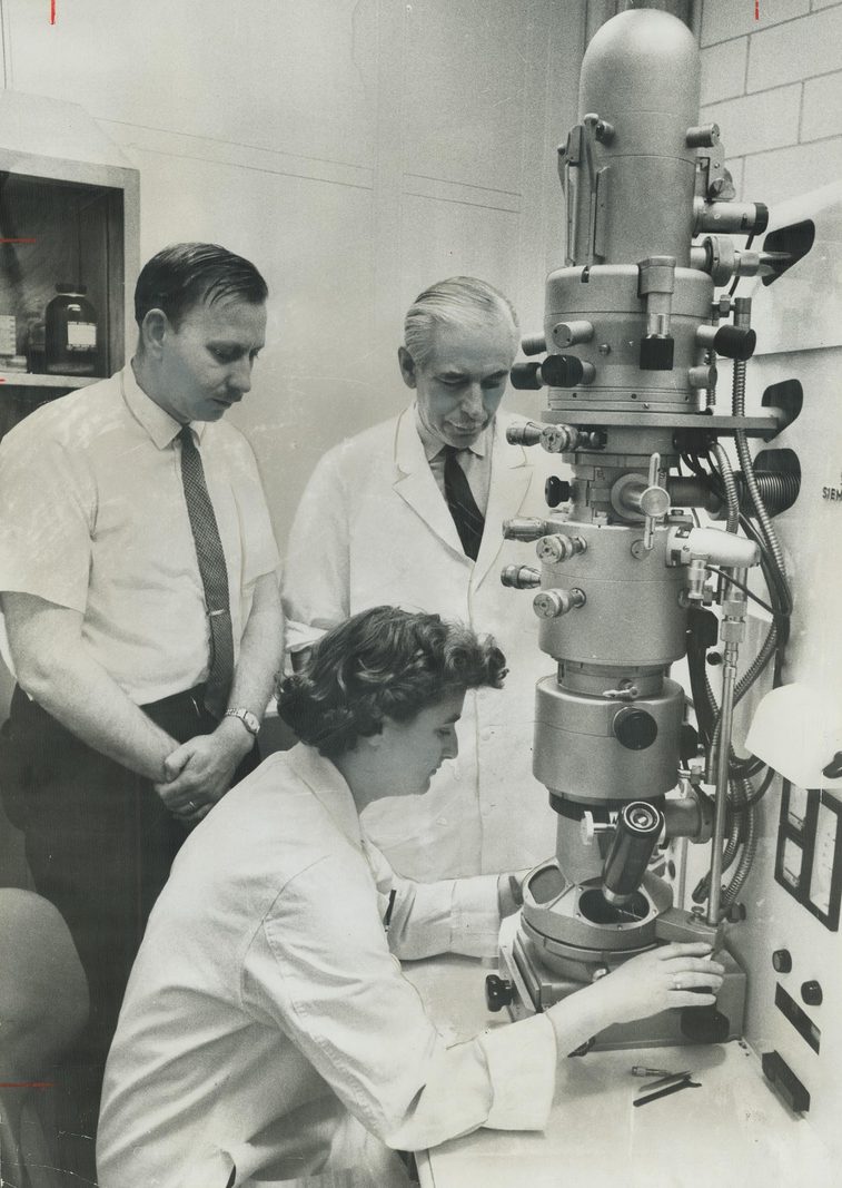
A laboratory education
Almeida left school at sixteen because university was beyond the family's means. In 1947 she began work in histopathology at Glasgow Royal Infirmary, then moved to the same field at St Bartholomew's in London. After emigrating to Canada, she found a position as an electron-microscopy technician at the Ontario Cancer Institute in Toronto. Joyce Almeida's obituary describes how publications and demonstrated ability opened a scientific career despite the missing university credentials.[3]
That route makes the word technician worth pausing over. Preparing a specimen determines what an observer can subsequently claim to have seen. The instrument supplies magnification; the preparation and the observer's judgment determine whether the enlarged object is informative. Almeida's papers make this usually hidden part of discovery unusually visible.
In March 1963, she and Allan Howatson published a method for applying negative staining to cell-associated virus. Their affiliation joined the Ontario Cancer Institute to the University of Toronto's Department of Medical Biophysics.[4] The paper places her development inside a working research group, with practical preparation methods themselves becoming publishable knowledge.
Giving an outline something to stand against
Negative staining creates contrast around biological material. Electron-dense stain surrounds particles and enters accessible spaces, helping their shapes stand out. A later methodological review in the CDC's Emerging Infectious Diseases explains the attraction: an observer can inspect a specimen for recognizable forms without first choosing an antibody against one suspected virus. The same review stresses preparation, particle concentration, and the limits of interpreting morphology.[5]
The distinction is consequential. A targeted test begins with a question about a particular agent. A broad visual examination can expose something outside that expectation, provided enough recognizable material reaches the grid. Its openness does not make it infallible. The observed shape still has to be judged, and a poor sample can leave little to judge.
This helps explain why Almeida's practical expertise mattered. A stronger microscope alone could not settle whether a faint or damaged particle represented an unfamiliar organism. Improving what survived preparation gave comparison a better starting point.
Antibodies could also make relationships visible
In a separate September 1963 study, Almeida, Bernhard Cinader, and Howatson examined negatively stained mixtures of viruses and antibodies. Their electron micrographs showed antibodies linking particles into aggregates, sometimes into rings. Under conditions of extreme antibody excess, the pattern changed: particles became heavily coated, and cross-linking was uncommon.[6]
That observation complicates the appealing idea that adding more reagent simply produces a better result. The appearance depended on the balance between the ingredients. An image could reveal a biological interaction, but it also recorded the conditions under which the interaction had been arranged.
This work supplied a way to combine visual form with immunological relationships. It also establishes an important boundary in Almeida's biography. Antibody-assisted imaging and ordinary negative staining were related tools in her repertoire. The coronavirus paper's methods used negative staining without adding antibodies.[1]
The common cold supplied a difficult specimen
A. P. Waterson recruited Almeida to St Thomas' Hospital Medical School in London in 1964 after meeting her in Toronto.[3] Meanwhile, at the Common Cold Research Unit near Salisbury, Tyrrell and M. L. Bynoe were confronting samples that resisted familiar culture methods.
Their June 1965 paper described B814, an agent associated with colds that could be propagated using organ cultures. These retained respiratory tissue rather than reducing the experiment to a conventional layer of cultured cells. Failure in standard cultures had left a gap between evidence that infectious material was present and the ability to characterize it.[7]
The practical lesson was already substantial: a laboratory's failure to grow an agent could reflect the growth conditions. The patient's illness did not have to conform to the laboratory's existing repertoire. Organ culture made different material available for examination; microscopy could then ask what that material contained.
Almeida and Tyrrell's 1967 study gently dispersed cells from organ cultures and examined negatively stained preparations. They checked known viruses and uninoculated cultures as part of the investigation. B814 and 229E resembled avian infectious bronchitis virus; a third agent, LAKEY, resembled the parainfluenza group.[1]
The third result matters: the method distinguished appearances, giving researchers grounds to keep one agent outside the proposed relationship.
A name followed the comparison
On 16 November 1968, a Nature notice reported a proposed name for the group: coronaviruses. Eight virologists, including Almeida and Tyrrell, connected the shared fringe of rounded surface projections to the appearance of the solar corona. Human and animal viruses could now be discussed together through a common morphological description.[8]
The notice recorded a naming proposal within an ongoing classification process. It should not be flattened into a claim that a modern virus family sprang fully formed from one laboratory encounter. Cultivation, imaging, comparison, and nomenclature were separate contributions, made by several researchers over several years.
Nor did the resemblance establish that all members would cause the same illness. A category organizes further questions; it does not answer every question about the organisms placed inside it. Reading the naming notice alongside the earlier experiments shows how cautiously a useful generalization could emerge.
Making the skill transferable
Almeida returned to the practical problem in her 1980 review, Practical aspects of diagnostic electron microscopy. She described negative staining's movement from research on viral structure into diagnostic work and set out the steps needed to obtain useful preparations.[9] The emphasis remained on making the method work in another person's hands.
That is an especially revealing endpoint for this part of her career. A celebrated image can circulate independently of its maker. A usable method carries something harder to preserve: the choices that made the image possible. Teaching those choices lets another laboratory reproduce the conditions for recognition.
Almeida's microhistory therefore lives at the meeting point of craft and interpretation. She helped make unfamiliar particles clear enough to compare, while experiments and collaborators supplied reasons to take the comparisons seriously. The enduring achievement was a way of looking that could be explained, checked, and passed on.
Sources
- June D. Almeida and D. A. J. Tyrrell, “The Morphology of Three Previously Uncharacterized Human Respiratory Viruses that Grow in Organ Culture,” Journal of General Virology 1:175–178 (1 April 1967) — original methods, controls, and distinctions among B814, 229E, and LAKEY.
- National Museums Scotland, “8 women in Scotland who shaped the history of science,” June Almeida section — provenance and credit for Norman James's 1963 photograph; used here for the photograph rather than the page's compressed discovery chronology.
- Joyce Almeida, “June Almeida (née Hart),” BMJ 336:1511 (28 June 2008) — obituary documenting her laboratory employment and recruitment to St Thomas'.
- June D. Almeida and Allan F. Howatson, “A Negative Staining Method for Cell-Associated Virus,” Journal of Cell Biology 16:616–620 (1 March 1963) — original method paper and Toronto institutional affiliation.
- Paul R. Hazelton and Hans R. Gelderblom, “Electron Microscopy for Rapid Diagnosis of Emerging Infectious Agents,” Emerging Infectious Diseases 9:294–303 (March 2003) — explanation of negative staining, visual identification, specimen quality, and diagnostic limits.
- June Almeida, Bernhard Cinader, and Allan Howatson, “The Structure of Antigen-Antibody Complexes: A Study by Electron Microscopy,” Journal of Experimental Medicine 118:327–340 (1 September 1963) — original observations of cross-linking and antibody excess.
- D. A. J. Tyrrell and M. L. Bynoe, “Cultivation of a Novel Type of Common-cold Virus in Organ Cultures,” BMJ 1:1467–1470 (5 June 1965) — original report on B814 and the culture problem.
- “Virology: Coronaviruses,” Nature 220:650 (16 November 1968) — contemporary notice of the proposed group name and its morphological rationale.
- J. D. Almeida, “Practical aspects of diagnostic electron microscopy,” Yale Journal of Biology and Medicine 53:5–18 (January–February 1980) — her account of specimen preparation and the method's diagnostic use.