A ball held inside a cage makes an unusually blunt substitute for the delicate flaps of a heart valve. Yet the Starr–Edwards design gave patients with severe valve disease a future that surgery had struggled to provide. A few years later, chemically treated pig valves offered another route to replacement.[1][3]
The two designs made different demands on the years after surgery. Mechanical valves promised endurance, accompanied by treatment to prevent clots. Tissue valves could reduce that medication burden, while leaving the possibility of another operation as the material deteriorated.[3][6] Their history explains why a device's working life and a patient's life could never be measured by the same clock.
Oregon: make the replacement keep moving
When engineer Lowell Edwards approached surgeon Albert Starr in Oregon in 1958, Edwards wanted to build an artificial heart. Starr proposed beginning with a valve. His 2007 recollection describes experiments in which early artificial leaflets moved successfully immediately after implantation in dogs, only to become obstructed by clot.[1]
That sequence matters: restoring motion at surgery did not establish that the implant could keep functioning in blood. The team moved toward a ball enclosed by a cage and repeatedly revised the surrounding structure. In human patients, anticoagulant treatment became part of the solution. Starr also committed to lifetime follow-up, recognizing that implantation began a continuing experiment.[1]
The first successful Starr–Edwards replacement followed in September 1960. The Lasker Foundation's account records that the recipient lived another ten years, dying after a fall from a ladder.[3] Such survival made the achievement tangible. It also opened a longer period in which complications could appear.
Paris: change what blood meets
Alain Carpentier approached the problem through a patient whose replacement valve had already saved his life. In his 2007 account, Carpentier recalled an artist who suffered a cerebral embolism after surgery and could no longer paint. He made reducing valve-related clot formation his research problem.[2]
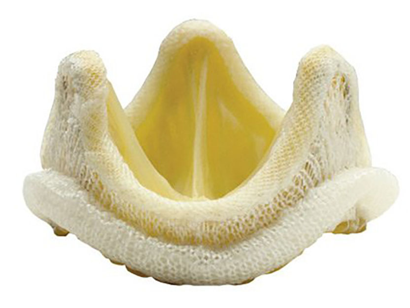
Animal tissue offered a promising blood-contacting surface, but preserving it was difficult. Early pig-valve implants developed inflammatory reactions and deterioration of collagen, the tissue's structural protein. In 1968, Carpentier identified how glutaraldehyde treatment could stabilize collagen and reduce immune reactions. The treated valve could then be mounted in a supporting frame.[2]
The resulting bioprosthesis was chemically preserved material with a biological origin. Its success depended on that material remaining serviceable; it was not a living replacement expected to renew itself. Later calcification and leaflet damage exposed the limit of preservation.[2]
The comparison had therefore become more interesting than engineering versus nature. Both approaches required engineering. One depended on an enduring mechanical structure and clot prevention; the other on preserving tissue while reducing its tendency to provoke clotting.
Edinburgh: survival and an intact valve parted company
The Edinburgh trial enrolled patients between 1975 and 1979. Its long-term analysis included 533 people, comparing a Björk–Shiley mechanical valve with Hancock or Carpentier–Edwards porcine valves. Its mechanical valve used a tilting disc, unlike the Starr–Edwards ball.[4]
The 2003 report found no statistically significant difference in overall survival at twenty years. Reoperation for valve failure, however, had occurred in 91 tissue-valve recipients and 22 mechanical-valve recipients. Major bleeding was more frequent with the mechanical valve. All mechanical-valve patients received warfarin; tissue-valve patients could also receive it at their cardiologist's discretion.[4]
These outcomes describe different experiences. Being alive after twenty years does not reveal whether someone needed another operation. Retaining an original valve does not reveal whether the person suffered a serious bleed. Overall survival leaves both questions open.
The American trial complicated the verdict
A separate Veterans Affairs trial studied 575 patients at 13 medical centers. Unlike Edinburgh's mixed-sex population, this was an all-male cohort. Its 2000 report found a survival advantage for mechanical valves in the aortic position: estimated all-cause mortality at fifteen years was 66% with mechanical valves versus 79% with tissue valves. It found no corresponding survival advantage for mitral replacement.[4][5]
Those are mortality estimates, not survival percentages. Keeping the endpoint attached to the number prevents an especially consequential misreading.
The VA trial also found more primary valve failure with tissue valves, particularly among younger patients, and more bleeding with mechanical valves.[5] The broad tradeoff resembled Edinburgh's; the survival finding was less uniform.
Together, the studies resist a universal winner. Valve position, the population studied, and the endpoint being counted all affect the conclusion. Their results concern particular devices and care delivered in that historical period. They cannot supply an unchanged forecast for someone receiving a different valve today.
A choice that extends beyond the operating room
The American Heart Association's patient guidance, reviewed in July 2026, still frames replacement around durability, medication needs, future procedures, and personal circumstances. Mechanical valves usually entail lifelong anticoagulation with monitoring. Tissue valves usually avoid that lifelong requirement, but may wear out and need another intervention.[6]
“Usually” carries real weight here. Avoiding a lifelong requirement imposed by the valve does not promise freedom from every future medication need. The whole person's treatment needs matter alongside the implanted material.
These histories suggest a more useful meaning of progress than finding a valve that wins every comparison. Starr's team made long survival possible with a manufactured mechanism. Carpentier's work expanded the ways patients could live with replacement. Long follow-up then revealed what each approach asked of them.
A valve must open and close reliably. The choice also concerns the years surrounding those movements: medication, monitoring, the possibility of bleeding, and the prospect of another procedure. Durability remains essential, but it has never been the only thing a patient has to live with.
Sources
- Albert Starr, “The artificial heart valve,” Nature Medicine (2007) — the surgeon's retrospective account of the Oregon experiments, anticoagulation, and commitment to lifetime follow-up; PDF hosted by the Lasker Foundation.
- Alain Carpentier, “The surprising rise of nonthrombogenic valvular surgery,” Nature Medicine (2007) — first-person account of tissue preservation, glutaraldehyde, and later material deterioration; Lasker Foundation PDF.
- Lasker Foundation, “Prosthetic aortic and mitral valves,” 2007 clinical research award — historical account, September 1960 implantation, and the paired valve photographs credited to Albert Starr and Edwards Lifesciences.
- H. Oxenham et al., “Twenty year comparison of a Bjork-Shiley mechanical heart valve with porcine bioprostheses,” Heart 89:715–721 (2003) — original randomized study, methods, outcomes, and comparison with the VA trial; publisher's PDF in the University of Edinburgh repository.
- Karl Hammermeister et al., final 15-year report of the Veterans Affairs randomized valve trial, Journal of the American College of Cardiology 36:1152–1158 (2000) — original comparison, valve-position differences, mortality, deterioration, and bleeding; PubMed abstract.
- American Heart Association, “Heart Valve Replacement: Types, Procedures and Treatment Options,” reviewed July 21, 2026 — patient guidance on durability, anticoagulation, future procedures, and individualized selection.