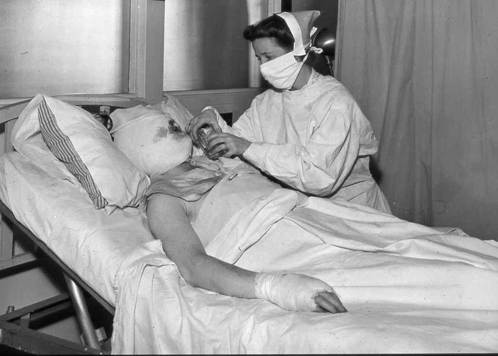
The nurse in the photograph is looking down at a bandage, but the patient’s injuries cannot be contained by the dressing. Smoke has entered lungs. Heat has altered skin and the circulation beneath it. Fluid is leaving the bloodstream for damaged tissue. Infection, stiffening joints, fear, and grief wait on different clocks. The bedside scene is quiet; the medicine demanded by it is not.[9]
That mismatch is the most important medical legacy of the Cocoanut Grove fire of November 28, 1942. The catastrophe is often credited with a tidy list of “firsts”—a new burn dressing, early penicillin, a fluid formula. The stronger reconstruction is about integration. In a few hours, Boston hospitals had to treat one disaster as an airway emergency, a shock state, a wound problem, a mass-casualty operation, and the beginning of prolonged rehabilitation.[8]
Boston’s official investigation counted 490 deaths and 166 injuries; later medical and institutional accounts use 491 or 492 deaths, so “nearly 500” is more honest than pretending the historical tallies are identical.[1][8][9] Massachusetts General Hospital received 114 casualties between about 10:30 p.m. and 12:45 a.m.; 75 were dead on arrival or died soon afterward, leaving 39 patients to be admitted and treated.[2] Those numbers compressed clinical judgment into minutes. They also exposed a fact that visible burns could hide: many of the gravest injuries were inside the airway or bloodstream.
The fire did not create modern burn care in one miraculous night. Wartime research on wound infection and burn physiology was already under way at Mass General, and the largest sustained survival gains came decades later with organized burn centers, dedicated teams, early wound closure, and critical-care specialization.[2][8] Cocoanut Grove mattered because it forced separate questions into the same room—and then made clinicians publish what they had learned.
November 28, 1942: the first problem was not a treatment
The fire moved through an overcrowded club with extraordinary speed. The Boston Fire Commissioner’s report placed about 1,000 people inside, estimated that flame reached the street-floor lobby in two to four minutes and crossed the main floor within five, and recorded the first civilian alarm at 10:20 p.m.[1] Smoke, flame, constricted exits, and the pressure of a fleeing crowd produced deaths before any hospital therapy could matter. The same report preserved a teenage busboy’s testimony about lighting a match near the first observed fire but formally ruled the origin unknown.[1] The distinction matters: a disaster begins as architecture, materials, crowd movement, and command before it becomes a diagnosis, and a memorable anecdote is not the same thing as a proven cause.
At Mass General, the first medical task was therefore sorting, not innovation. The emergency ward received bodies and living patients in the same surge. The hospital administrator’s 1943 account says nearly the entire emergency organization was assembled by 11:15 p.m. and all 39 living patients were in beds, with initial dressing and treatment under way, by 1:30 a.m.[2] Some had obvious surface burns; others carried respiratory damage whose severity was harder to read at first glance.
That distinction reorganized the event. If “burn victim” meant only a person with damaged skin, the clinical picture made little sense. Some people with limited visible burning were critically ill. Others who survived the initial smoke and heat still faced shock, delayed lung injury, wound infection, and loss of movement. The intake forced clinicians to stop treating the skin as a complete account of the injury.
The airway made the hidden burn visible
The primary clinical report supplied some of the clearest evidence. Only three of the 39 living Mass General patients were wholly free of respiratory symptoms, and the severity of the surface burn correlated poorly with the severity of respiratory damage.[3] An early wave of breathing difficulty appeared around three hours after the fire. The most dangerous delayed phase began around 24 hours and continued for roughly another 36.[3]
The time lag mattered. Carbon monoxide could kill rapidly, while thermal and chemical injury to the respiratory tract could evolve after a patient had escaped the building. A face might be burned without deep airway injury, or an airway might be gravely injured without a large surface wound. Clinicians had to join the circumstances of exposure—an enclosed, smoke-filled room—to voice changes, breathing difficulty, oxygenation, secretions, radiographs, and later pathology. All seven Mass General patients who died after admission were judged to have died from pulmonary complications.[2][3]
The June 1943 Mass General symposium consequently devoted separate papers to immediate resuscitation, pulmonary lesions, clinical respiratory findings, and the combined problem of burn shock with lung damage.[3][5][8] That division of labor was itself an advance. “The burn” no longer named one surface. It named interacting injuries that different specialists had to describe together.
Shock turned burn area into a fluid question
The second hidden injury was circulatory. A major burn does not simply remove skin. It changes capillary permeability and pulls fluid out of the vascular space into injured tissue. Blood pressure and urine output can deteriorate even when external bleeding is not the central problem. In 1942, clinicians understood parts of this process, but the amount and timing of replacement fluid were not yet governed by the standardized protocols familiar to later burn units.[2][5]
Cocoanut Grove supplied a brutally concentrated clinical series. Oliver Cope and Frederic Rhinelander’s 1943 paper treated burn shock and pulmonary damage as one combined problem, not two independent diagnoses.[5] Their historical starting plan called for 500 cubic centimeters of plasma per 10% of body surface burned over 24 hours, initially accompanied by an equal volume of saline, with repeated hematocrit and serum-protein measurements used to revise treatment.[5] This is a reconstruction of 1942 practice, not a current dosing instruction. Its importance lies in the feedback logic: resuscitation had to be estimated, observed, and changed.
The later “burn budget” work associated with Cope and Francis Moore related fluid needs to the extent of the burn and the patient’s body size. Their 1947 paper also acknowledged that a National Research Council conference had recommended surface-area-guided plasma therapy in January 1942, months before the fire.[7] Cocoanut Grove therefore refined and stress-tested an emerging approach; it did not conjure fluid resuscitation from nothing. The line of work helped lead toward later formulas, but it should not be collapsed into the modern Parkland formula or treated as a frozen recipe discovered on fire night.[7][8] The durable change was more basic: surface area became a physiologic variable, and fluid became a dose to be adjusted against the patient’s response rather than an undifferentiated act of rescue.
This was one reason measurement of the wound mattered. Mapping the percentage of body surface burned was not clerical description. It connected what clinicians could see on the skin to what they needed to anticipate in the circulation.
The dressing became gentler, but care became more active
Surface treatment was changing at the same time. Before the fire, one common approach used tannic acid and dyes to “tan” the wound into a hard eschar. Mass General’s later review characterizes the pre-1943 era as one of tanning methods, spontaneous eschar separation, limited intravenous therapy, and systemic antibiotics largely restricted to sulfa drugs.[8]
Cope’s paper on the 39 treated patients described a softer wound method built around minimal preparatory handling—no routine cleansing or mechanical debridement—followed by boric-petrolatum gauze and bulky dry dressings rather than chemical tanning.[4] First dressings generally remained in place for five to ten days; deeper wounds were prepared for grafting later, not immediately excised at the bedside.[4] The visual difference could look modest—one bandage replacing another—but its logic was not. The team was trying to protect viable tissue, make the wound observable, reduce additional injury from treatment, and preserve a path toward healing or grafting.
The 1943 symposium placed wound care beside hand rehabilitation and physical therapy.[8] That sequence matters. Survival was not the only outcome, especially for burns across hands, faces, and joints. A closed wound could still leave a patient unable to move, work, or care for themselves. The medical unit of success was beginning to stretch from the first dressing toward function.
The cover photograph catches that transition without romanticizing it. The nurse’s mask, gown, jar, gauze, and the patient’s wrapped head and hand show burn care as repeated bedside labor. No single object in the frame is “the breakthrough.” The treatment is the coordinated attention around the patient.[9]
Penicillin entered as evidence with a warning label
Antimicrobial treatment has acquired its own Cocoanut Grove legend. Penicillin was not the initial antibiotic. The 39 admitted patients first received sulfadiazine; penicillin began on day six, after fever and elevated white-cell counts appeared, and 13 patients received it at some point.[6] The main 11-patient group received 5,000 units intramuscularly every four hours—an early, cautious dose used while clinicians were still learning the drug’s safety and behavior.[6]
The episode was historically important, but it was not a clean trial of a miracle cure. Lyons’s original report called an accurate appraisal of penicillin’s efficacy impossible: the dose was later understood to be too small, and the drug was usually given with sulfadiazine.[6] Infection control also depended on wound handling, nursing, dressings, isolation practice, drainage, nutrition, and later graft care. Penicillin added a powerful tool; it did not turn the rest of the system into scenery.
That boundary makes the episode more useful, not less. Emergency medicine often produces evidence bundles before it produces controlled comparisons. Clinicians act with the best available rationale, observe closely, and then have to resist giving one component credit for everything that followed.
June 1943: a hospital response becomes a shared record
Seven months after the fire, Annals of Surgery published a Mass General symposium containing 16 articles on the response. Its range was remarkable: disaster organization, acute grief, airway management, radiology, pulmonary pathology, surface burns, infection, rehabilitation, physical therapy, burn shock, the blood bank, and metabolic change.[8]
The breadth reveals what Cocoanut Grove changed most decisively. The event did not yield one doctrine. It produced a common case record that made surgeons, anesthetists, pathologists, radiologists, bacteriologists, nurses, rehabilitation staff, social workers, and psychiatrists legible to one another. The patient moved through all of their domains, so the published explanation had to move through them too.
There was a wartime channel ready to carry the findings. The U.S. Office of Scientific Research and Development had already supported Mass General work on compound-wound infection and burn physiology. Publication in 1943 gave the disaster’s lessons an audience far beyond Boston at a moment when military services were preparing to treat large numbers of burns and contaminated wounds.[2][8]
What Cocoanut Grove changed—and what it did not
A high-quality origin story needs a stopping rule. The fire’s response generated durable concepts, but it did not instantly deliver the survival rates of a modern burn center. Mass General’s 70-year review found that mortality declined from 15.6% in 1939–1942 to 10.5% in 1943–1954, but the difference was only a borderline statistical trend and did not persist as a clear long-term leap. At that institution, the largest sustained improvement arrived in the 1970s, when specialized burn centers joined organized critical care, dedicated multidisciplinary teams, early excision and closure, and specialist support.[8]
That later evidence corrects the heroic version without shrinking the event. Cocoanut Grove’s medical importance lies less in a claim that one night “invented” burn medicine than in the way it redrew the problem. Airway injury could be lethal without dramatic skin burns. Fluid loss had to be related to body and wound. A dressing could harm as well as protect. Antibiotics had to be evaluated inside a treatment bundle. Rehabilitation began before the wound story was over. Grief belonged in the clinical record.
Return, finally, to the nurse and patient. The photograph is not an illustration of a single discovery. It is a portrait of medicine learning to hold several clocks at once: minutes for airway and shock, days for lung injury and infection, weeks for closure, months for movement, and years for memory. The burn extended beyond the skin, so care had to do the same.
Sources
- William Arthur Reilly, Report Concerning the Cocoanut Grove Fire, November 28, 1942 (Boston Fire Department, submitted November 19, 1943) — official investigation used for the crowd estimate, fire timeline, casualty count, match testimony, and unknown-cause finding.
- Nathaniel W. Faxon, “The Problems of the Hospital Administration,” Annals of Surgery 117 (1943) — primary Mass General account of the 114 arrivals, 39 living patients, emergency mobilization, initial treatment sequence, and pre-existing research program.
- Joseph C. Aub, Howard Pittman, and Austin M. Brues, “The Pulmonary Complications: A Clinical Description,” Annals of Surgery 117 (1943) — primary report on respiratory symptoms, their weak relation to surface-burn severity, delayed deterioration, and pulmonary deaths.
- Oliver Cope, “The Treatment of the Surface Burns,” Annals of Surgery 117 (1943) — primary report on the 39-patient soft-treatment regimen, dressing interval, treatment uncertainties, and later preparation of deep burns for grafting.
- Oliver Cope and Frederic W. Rhinelander, “The Problem of Burn Shock Complicated by Pulmonary Damage,” Annals of Surgery 117 (1943) — primary report on the historical resuscitation plan, laboratory feedback, and the interaction between fluid therapy and lung injury.
- Champ Lyons, “Problems of Infection and Chemotherapy,” Annals of Surgery 117 (1943) — primary report on initial sulfadiazine, day-six penicillin use, concurrent therapy, dose limitation, and the authors’ inability to isolate efficacy.
- Oliver Cope and Francis D. Moore, “The Redistribution of Body Water and the Fluid Therapy of the Burned Patient,” Annals of Surgery 126 (1947) — follow-on fluid study and formula, including its account of pre-fire surface-area-guided recommendations.
- Ronald G. Tompkins, “Survival from Burns in the New Millennium: 70 Years Experience from a Single Institution,” Annals of Surgery 261 (2015) — retrospective synthesis of the 16-paper symposium, treatment changes, mortality trend, and the later organized-burn-center survival leap.
- Mass General Research Institute, “How the Cocoanut Grove Fire Changed Burn Care at Mass General and Beyond” (November 28, 2022) — institutional history and MGH Archives source for the bedside-care photograph used as the article image.