A ventilator can keep delivering the same breaths while the carbon dioxide measured at the airway falls. The change may begin in the circulation: less blood reaches the lungs, carrying less CO₂ for those breaths to remove. A monitor beside the breathing tube can therefore register a problem upstream of breathing itself.[3]
That is the useful complication inside capnography. Its moving trace records carbon dioxide in respiratory gas. Understanding the trace means following the gas through the body before it reaches the sensor.
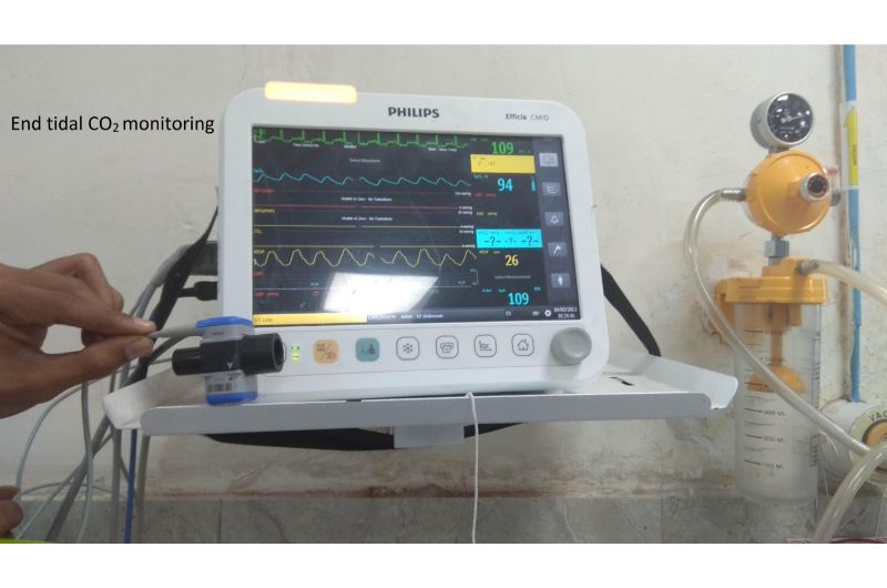
The journey ends at the sensor
Cells produce CO₂ during metabolism. Blood transports it to the lungs; there it passes into the air sacs, or alveoli, and leaves with exhalation. Production, pulmonary blood flow and alveolar ventilation all affect what emerges. The capnograph observes the end of that journey.[1]
Its waveform also preserves the order in which a breath empties. Gas from the conducting airways arrives first, carrying little CO₂ because those airways do not exchange gas with blood. Gas from the alveoli follows, raising the trace toward a plateau. The end-expiratory reading is called end-tidal CO₂, usually abbreviated EtCO₂. The next inspiration brings the trace back toward baseline.[1]
A mainstream instrument measures gas at an adapter in the breathing circuit. A sidestream instrument draws a sample through tubing to an analyzer farther away. Both arrangements connect the display to a physical sample; neither measures blood directly.[4]
Hold breathing steady, and circulation becomes visible
A November 1994 study by K. Shibutani and colleagues examined this connection during abdominal aortic aneurysm surgery. The investigators made 33 measurements in 24 patients, keeping ventilation constant and measuring soon after changes in circulation. Percentage falls in end-tidal CO₂ tracked percentage falls in cardiac output, the volume of blood the heart pumps per minute, with R² = 0.82.[3]
The point is the condition attached to the finding. Because ventilation remained fixed, a fall in exhaled CO₂ could reveal reduced delivery of the gas to the lungs. The correlation describes that study's observations; it does not provide a universal formula for calculating cardiac output from a capnograph.
A second experiment, published on March 26, 2012, approached the connection from the other direction. Manuel Ignacio Monge García and colleagues studied 37 mechanically ventilated patients with acute circulatory failure. A brief passive leg raise shifted blood toward the chest. Changes in cardiac output and end-tidal CO₂ were strongly associated, with R² = 0.79.[5]
Here too, ventilation was held fixed, and the researchers assumed tissue CO₂ production remained stable. They proposed two contributing mechanisms: increased blood flow delivered more CO₂, and better pulmonary perfusion could bring more ventilated air sacs into effective gas exchange. They had not measured arterial CO₂, so they could not separate those contributions. The authors also warned against extending their results to patients breathing spontaneously.[5]
These experiments make the same interpretive demand: before assigning a change in the trace to one cause, establish what else was changing.
The airway and the artery can disagree
Some air reaches lung regions that receive too little blood for effective gas exchange. This contributes to physiological dead space: ventilation that does little useful exchange. As dead space increases, the difference between arterial CO₂ and end-tidal CO₂ can widen.[1]
The American Association for Respiratory Care's April 2011 guideline makes that boundary explicit. End-tidal monitoring supplies information about ventilation and perfusion, but it cannot replace assessment of arterial CO₂. The difference can vary within the same patient over time.[4]
A small number at the airway therefore does not establish a small CO₂ burden in arterial blood. Treating the two measurements as interchangeable removes precisely the circulation and gas-exchange information that makes capnography useful.
The sample has its own vulnerabilities
The gas must also reach the instrument. Condensation, secretions or obstruction in a sampling system can make its result unreliable.[4] In patients wearing a nasal sampling cannula, displacement and dilution can complicate interpretation.[1] The Anesthesia Patient Safety Foundation explains that capnography in non-intubated patients often provides more dependable information about respiratory timing than about the exact arterial CO₂ level.[6]
A lost trace demands attention. It can reflect a patient or airway problem, but a blocked sampling line can also erase the waveform. The display alone cannot locate the failure.[1] Clinical assessment remains part of interpreting the signal.[8]
Oxygen answers another part of the question
Oxygen saturation supplies essential information alongside this CO₂ story. A 2004 controlled study by Eugene S. Fu and colleagues showed why it cannot stand in for ventilation: supplemental oxygen maintained saturation readings during deliberately reduced ventilation, while arterial and end-tidal CO₂ rose.[7]
The oxygen was doing something real. Preserved oxygenation simply did not demonstrate adequate CO₂ clearance. That distinction explains the need for complementary measurements without making either instrument redundant.
The American Society of Anesthesiologists' monitoring standards, amended on October 15, 2025, separately address oxygenation, ventilation and circulation. For an inserted endotracheal tube or supraglottic airway, they call for clinical verification together with identification of exhaled CO₂, followed by ongoing end-tidal analysis through the specified period of airway use.[8]
Read that separation back into the bedside screen. Each breath carries information about gas movement, but also about the blood that supplied the gas. A capnograph becomes most informative when the clinician follows that whole route.
Sources
- Haley Perkins and Debnath Chatterjee, “Capnography,” OpenAnesthesia (updated March 10, 2026) — physiology, waveform phases and measurement principles.
- Dr. D. Y. Patil Medical College, Hospital and Research Centre, Department of Emergency Medicine — original photograph of end-tidal CO₂ monitoring equipment.
- K. Shibutani et al., “Do changes in end-tidal PCO₂ quantitatively reflect changes in cardiac output?” Anesthesia & Analgesia 79:829–833 (November 1994) — original study abstract.
- Brian K. Walsh, David N. Crotwell and Ruben D. Restrepo, “Capnography/Capnometry During Mechanical Ventilation: 2011,” AARC Clinical Practice Guideline, Respiratory Care 56:503–509 (April 2011) — sampling configurations and limitations, especially section 7.
- Manuel Ignacio Monge García et al., “Non-invasive assessment of fluid responsiveness by changes in partial end-tidal CO₂ pressure during a passive leg-raising maneuver,” Annals of Intensive Care 2:9 (March 26, 2012) — original study and its physiological assumptions.
- Rajnish K. Gupta and David A. Edwards, “Monitoring for Opioid-Induced Respiratory Depression,” APSF Newsletter (February 2018), pages 70–71 — interpretation and limitations of monitoring in non-intubated patients.
- Eugene S. Fu et al., “Supplemental Oxygen Impairs Detection of Hypoventilation by Pulse Oximetry,” CHEST 126:1552–1558 (November 2004) — original paper hosted by the University of Florida.
- American Society of Anesthesiologists, “Standards for Basic Anesthetic Monitoring” (amended October 15, 2025) — separate monitoring requirements for oxygenation, ventilation and circulation.